Miscellaneous Arthritides and Arthropathies


Miscellaneous Arthritides and Arthropathies


Connective Tissue Arthropathies

An overview of the clinical and radiographic hallmarks of the forms of arthritis associated with connective tissue disorders is presented in Table 15.1.


Systemic Lupus Erythematosus

Systemic lupus erythematosus (SLE) is a chronic, inflammatory, connective tissue disorder of unknown cause characterized by significant immunologic abnormalities and involvement of multiple organs. The risk of developing this condition is in part genetic, but it is a complex genetic disorder with no clear Mendelian pattern of inheritance, although it occurs in families. The first described genetic link to SLE was the major histocompatibility complex (MHC) on chromosome 6, which contains the human lymphocytic antigens (HLA)-DR. More recent investigations concluded that programmed cell death 1 (PDCD1) gene is responsible for the linkage at chromosome 2q34 and is associated with lupus nephritis. Women, particularly adolescents and young adults, are affected nine times as frequently as men, and this prevalence may be linked to TLR7 gene. The clinical manifestations of SLE vary according to the distribution and extent of systemic alterations. The most common symptoms are malaise, weakness, fever, anorexia, and weight loss. Consistent and characteristic features of this disease are serologic abnormalities, including a variety of serum autoantibodies to nuclear antigens, which have been historically associated with the presence of lupus erythematosus cells and neutrophilic leukocytes filled with cytoplasmic inclusion bodies.

Antinuclear antibodies (ANAs) are useful in the differential diagnosis of SLE, and changes in the titer of antibodies to DNA are useful in following disease activity. ANAs are a heterogeneous group of antibodies directed against a number of discrete nuclear macromolecular proteins. They represent what has classically been referred to as autoantibodies because they are directed against components normally present in all nucleated cells. They generally lack tissue or species specificity; therefore, they will cross-react with nuclei from different sources. The primary sources for study of these antibodies are patients with SLE and related systemic rheumatic diseases. Many studies have centered on defining the specificity of these antibodies and have contributed extensively to our understanding of their immunopathologic role in connective tissue disorders.

The musculoskeletal system is a frequent site of involvement in SLE, and joint abnormalities, exhibited by 90% of patients during the course of the disease, represent a significant part of the clinical and radiologic picture. Arthritic involvement is symmetric, and articular deformities without fixed contractures are a hallmark of this disorder. The hands are the predominant site of involvement. Typically, the conventional radiography discloses malalignments, most commonly at the metacarpophalangeal and proximal interphalangeal joints of the fingers and the first carpometacarpal, metacarpophalangeal, and the interphalangeal joints of the thumb (Fig. 15.1). These abnormalities may not be apparent on a dorsovolar radiograph because the malalignments are flexible and are corrected by the pressure of the hand against the radiographic cassette (Fig. 15.2). These pathognomonic deformities usually occur secondary to a loss of support from the ligamentous and capsular structures about the joint and, at least in the early stage of disease, are completely reducible. Only very seldom are these abnormalities fixed and/or accompanied by articular erosions (Fig. 15.3).

Some patients present with sclerosis of the distal phalanges (acral sclerosis) (Fig. 15.4) or with resorption of the terminal tufts (acroosteolysis). Osteonecrosis, which is frequently seen, has been attributed to complications of treatment with corticosteroids (Fig. 15.5). However, current investigations suggest the vital role of the inflammatory process (vasculitis) in the development of this complication.

The treatment of lupus consists of using nonsteroidal antiinflammatory drugs (NSAIDs), antimalarial drugs (such as hydroxychloroquine), corticosteroids (such as prednisone), and immunosuppressants (such as cyclophosphamide, azathioprine, mycophenolate, leflunomide, and methotrexate).


Scleroderma

Scleroderma (progressive systemic sclerosis) is a generalized disorder of unknown cause. It is seen predominantly in young women, usually becoming apparent in their third and fourth decades. Primarily a connective tissue disorder characterized by deposition of collagen and other components of extracellular matrix in the skin and internal organs, it is distinguished by thickening and fibrosis of the skin and subcutaneous tissues, with frequent involvement of the musculoskeletal system. Recently, researchers have identified a new genetic link to systemic form of scleroderma, a susceptibility locus involving the genome CD247 (that encodes the T-cell receptor zeta subunit modulating T-cell activation), in addition to previously known genes MHC, IRF5, and STAT4 (that encodes a regulatory protein important to immune system). Clinically, many patients develop joint involvement, which is manifesting as arthralgia and arthritis leading to flexion contractions of the fingers. Most patients have the so-called CREST syndrome, which refers to the coexistence of calcinosis, Raynaud phenomenon (episodes of intermittent pallor of the fingers and toes on exposure to cold, secondary to vasoconstriction of the small blood vessels), esophageal abnormalities (dilatation and hypoperistalsis), sclerodactyly, and telangiectasia; 30% to 40% of patients have a positive serologic test for rheumatoid factor and a positive ANA test.


TABLE 15.1 Clinical and Imaging Hallmarks of Connective Tissue Arthritides (Arthropathies)


Type of Arthritis


Site


Crucial Abnormalities


Technique/Projection


SLE (F > M; young adults; blacks > whites; skin changes: rash)


Hands


Hips, ankles, shoulders


Flexible joint contractures


Osteonecrosis


Lateral view


Standard views of affected joints


Scintigraphy


MRI


Scleroderma (F > M; skin changes: edema, thickening)


Hands


Soft-tissue calcifications


Acroosteolysis


Tapering of distal phalanges


Interphalangeal destructive changes


Dorsovolar and lateral views


Gastrointestinal tract


Dilatation of esophagus


Decreased peristalsis


Dilatation of duodenum and small bowel


Esophagram


Esophagram (cine or video study)


Upper gastrointestinal and small bowel series


Polymyositis/Dermatomyositis


Pseudodiverticulosis of colon


Barium enema


Upper and lower extremities (proximal parts)


Soft-tissue calcifications


Periarticular osteoporosis


Xeroradiography; digital radiography


Hands


Erosions and destructive changes in distal interphalangeal articulations


Dorsovolar and lateral views


MCTD (overlap of clinical features of SLE, scleroderma, dermatomyositis, and rheumatoid arthritis)


Hands, wrists


Erosions and destructive changes in proximal interphalangeal, metacarpophalangeal, radiocarpal and midcarpal articulations, associated with joint space narrowing


Dorsovolar and lateral views


Symmetric soft-tissue swelling


MRI


Soft-tissue atrophy and calcifications


Posteroanterior and lateral views


Chest


Pleural and pericardial effusions


Ultrasound


SLE, systemic lupus erythematosus; F, female; M, male; MRI, magnetic resonance imaging; MCTD, mixed connective tissue disease.


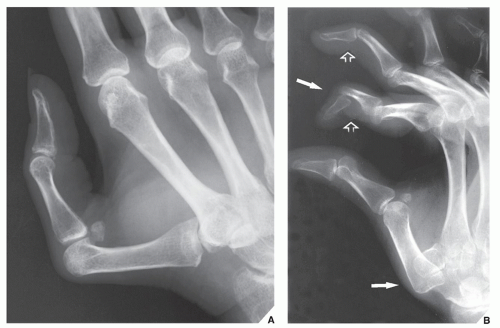
FIGURE 15.1 Systemic lupus erythematosus. (A) Typical appearance of the thumb in a 43-year-old woman with SLE. Note subluxations in the first carpometacarpal and metacarpophalangeal joints without articular erosions. (B) In another patient, a 32-year-old woman, the oblique radiograph of her left hand shows dislocations at the first carpometacarpal joint and distal interphalangeal joint of the index finger (arrows), and subluxations in the metacarpophalangeal joints of the index and middle fingers associated with swan-neck deformities (open arrows).


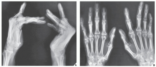
FIGURE 15.2 Systemic lupus erythematosus. (A) Lateral radiograph of both hands of a 42-year-old woman with documented SLE for the past 4 years demonstrates flexion deformities in the metacarpophalangeal joints. On the dorsovolar projection (B), the flexion deformities have been corrected by the pressure of the hands against the radiographic cassette.


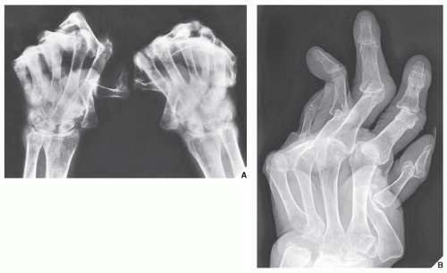
FIGURE 15.3 Systemic lupus erythematosus. (A) 62-year-old woman presented with a 15-year history of SLE. Dorsovolar view of both hands shows severe deformities, subluxations, and articular erosions. Note the advanced osteoporosis secondary to disuse of the extremities and treatment with corticosteroids. (B) In another patient, a 51-year-old woman, note flexion contractions, subluxations, and dislocations in the several joints of the right hand.


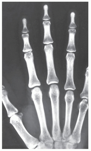
FIGURE 15.4 Systemic lupus erythematosus. Dorsovolar film of the hand of a 29-year-old woman with SLE demonstrates sclerosis of the distal phalanges (acral sclerosis). Similar sclerotic changes are also occasionally seen in rheumatoid arthritis and scleroderma.

Radiographically, scleroderma presents with characteristic abnormalities of the bone and soft tissues. The hands usually exhibit atrophy of the soft tissues at the tips of the fingers (Fig. 15.6), resorption of the distal phalanges (acroosteolysis), osteopenia, subcutaneous and periarticular calcifications (Figs. 15.7 and 15.8A), and destructive changes of the small articulations, usually the interphalangeal joints (Fig. 15.9). Soft-tissue calcifications within the upper limbs can occasionally be quite prominent (see Fig. 15.8B). Corroborative findings are seen in the gastrointestinal tract, where dilatation of the esophagus and small bowel, together with a pseudoobstruction pattern, is characteristic (Fig. 15.10). Pseudodiverticula in the colon are also commonly seen.

The treatment of scleroderma includes the use of antiinflammatory drugs such as NSAIDs, corticosteroids such as prednisone, immunosuppressive therapy with high dose of cyclophosphamide, anti-interferon agents such as sifalimumab, and anti-B-cell agents such as rituximab. Recent trials using autologous bone marrow transplantation yielded promising results.


Polymyositis and Dermatomyositis

Polymyositis and dermatomyositis are disorders of striated muscle and skin and are characterized by diffuse, nonsuppurative inflammation as well as degeneration. Early diagnosis and subsequent management of patients with any type of myopathy, including polymyositis and dermatomyositis, can be facilitated by the use of appropriate laboratory tests. The four tests most helpful in evaluating muscle disorders include (1) serum enzymes, (2) urinary creatine and creatinine excretion, (3) electromyogram, and (4) muscle biopsy.

Different serum enzyme determinations have been advocated, but the most valuable tests include serum creatine phosphokinase (CPK), serum aldolase (ALD), serum lactate dehydrogenase (LDH), serum glutamic oxalacetic transaminase (SGOT), and serum glutamic pyruvic transaminase (SGPT). Further, the determination of serum enzyme levels and urinary creatine excretion is helpful for the clinical management of polymyositis

and dermatomyositis because the two tests provide a broader perspective than either test alone.


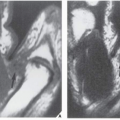
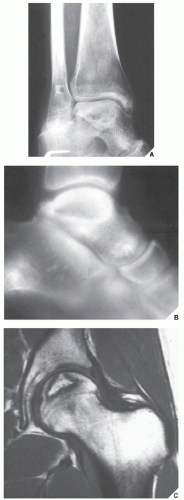
FIGURE 15.5 SLE complicated by osteonecrosis. Oblique radiograph (A) and lateral tomogram (B) of the ankle demonstrate osteonecrosis of the talus in a 26-year-old woman with lupus who was treated with massive doses of steroids. (C) Coronal T2-weighted MRI in an 18-year-old woman with SLE demonstrates a focal area of osteonecrosis of the femoral head.


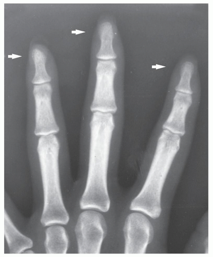
FIGURE 15.6 Scleroderma. A 24-year-old woman presented with atrophy of the soft tissues at the distal phalanges of the index, middle, and ring fingers (arrows).


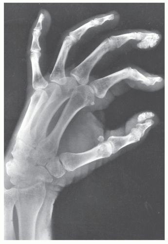
FIGURE 15.7 Scleroderma. A 32-year-old woman with progressive systemic sclerosis exhibits soft-tissue calcifications in the distal phalanges of the right hand (arrows), a typical feature of this disorder.


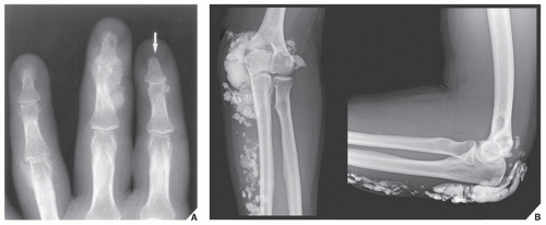
FIGURE 15.8 Scleroderma. (A) Dorsovolar radiograph of the fingers of a 44-year-old woman reveals acroosteolysis (arrow), soft-tissue calcifications, and destructive changes of the distal interphalangeal joint of the middle finger. (B) In another patient, a 46-year-old woman, extensive soft-tissue calcifications are present around the elbow and the forearm.


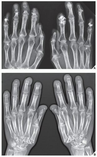
FIGURE 15.9 Scleroderma. (A) Dorsovolar radiograph of the hands of a 50-year-old man with documented systemic sclerosis shows destructive changes in the distal interphalangeal joints as well as soft-tissue calcifications and resorption of the tip of the distal phalanx of the left middle finger. (B) Dorsovolar radiograph of the hands of a 53-year-old woman with longstanding systemic sclerosis shows acroosteolysis of all distal phalanges. Note also erosions of the first carpometacarpal joints.

A positive biopsy may not only demonstrate that the disease process is myopathic, thus enabling the physician to rule out a neurogenic lower motor neuron lesion, but may also identify those patients whose muscle disease is more severe pathologically than was suspected on clinical grounds. This is important with respect to prognosis. With the aid of histochemical and electron microscopic techniques, muscle biopsy will occasionally enable the pathologist to diagnose one of the rare forms of myopathy that can clinically mimic polymyositis. Such diseases include sarcoid myopathy, central core disease, and muscle diseases associated with abnormal mitochondria.

The pathologic changes found on muscle biopsy in polymyositis have been well described. The degree of pathologic change may vary widely; one patient may show only negligible pathologic changes in muscle fibers on biopsy results, whereas another patient presenting similar clinical features may show extensive necrosis and fiber replacement. This variability in histologic findings is probably responsible for the frequent normal muscle biopsy results from patients with otherwise classic polymyositis. The overall rate of positive findings from muscle biopsy in several studies of polymyositis was in the range of 55% to 80%.

Imaging abnormalities in polymyositis and dermatomyositis are divided into two types: those involving soft tissues and those involving joints. The most characteristic soft-tissue abnormality in both conditions is soft-tissue calcifications. The favorite sites of intermuscular calcification are the large muscles in the proximal parts of upper and lower extremities. In addition, subcutaneous calcifications similar to those of scleroderma are seen (Fig. 15.11).

Articular abnormalities are rare. The most frequently reported, however, is periarticular osteoporosis. Destructive joint changes have been reported only occasionally and primarily in the distal interphalangeal articulations of the hands.


Mixed Connective Tissue Disease

Mixed connective tissue disease (MCTD) was first reported as a distinctive syndrome by Sharp and associates in 1972. This syndrome is characterized by clinical abnormalities that combine the features of SLE, scleroderma, dermatomyositis, and rheumatoid arthritis. The one feature that distinguishes MCTD as a separate entity is a positive serologic test for antibody to the ribonucleoprotein (RNP) component of extractable nuclear antigen (ENA). The typical clinical pattern consists of Raynaud
phenomenon, polyarthralgia, swelling of the hands, esophageal hypomotility, inflammatory myopathy, and pulmonary disease. Women constitute approximately 80% of affected patients. Patients with MCTD have prominent joint abnormalities, with typical involvement of the small articulations of the hand, wrist, and foot; large joints such as the knee, elbow, and shoulder may also be affected. The joint deformities mimic those seen in rheumatoid arthritis but occasionally joint subluxation may be nonerosive, as in SLE. Soft-tissue abnormalities are identical to those encountered in scleroderma (Figs. 15.12, 15.13, 15.14).


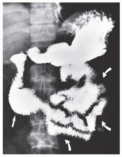
FIGURE 15.10 Scleroderma. Upper gastrointestinal series and small bowel study in the patient shown in Figure 15.9A demonstrate dilatation of the second and third portions of the duodenum (arrow) and jejunum (curved arrows), with a pseudoobstruction pattern.


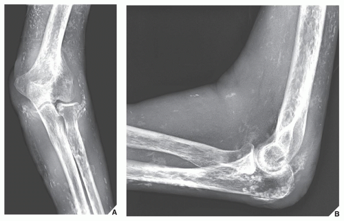
FIGURE 15.11 Dermatomyositis. (A) External oblique and (B) lateral radiographs of the left elbow of a 64-year-old woman show extensive soft-tissue calcifications, characteristic for this disorder. Note also prominent periarticular osteoporosis.


Vasculitis

There is a diverse clinical spectrum of the vasculitides that includes systemic necrotizing vasculitis, hypersensitivity vasculitis, Wegener granulomatosis, lymphomatoid granulomatosis, giant cell arteritis, and a variety of miscellaneous syndromes (e.g., Kawasaki disease, Behçet disease, and others). A discussion of these diverse but often overlapping diseases is far beyond the scope of this volume, but the reader is referred to several key references at the end of this chapter. The demonstration of vasculitis by angiograms can often be documented by the presence of aneurysmal dilatation in affected vessels. Generally, an angiogram is performed when the diagnosis cannot be established by tissue biopsy.


Metabolic and Endocrine Arthritides

An overview of the clinical and radiographic hallmarks of the arthritides associated with metabolic and endocrine abnormalities is shown in Table 15.2.


Gout

Gout is a metabolic disorder characterized by recurrent episodes of arthritis associated with the presence of monosodium urate monohydrate crystals in the synovial fluid leukocytes and, in many cases, gross deposits of sodium urate (tophi) in periarticular soft tissues. Serum uric acid concentrations are elevated.

The great toe is the most common site of involvement in gouty arthritis; the condition known as podagra, which involves the first metatarsophalangeal joint, occurs in approximately 75% of patients. Other frequently affected sites include the ankle, knee, elbow, and wrist. Most patients are men, but gouty arthritis is seen in postmenopausal women as well. Recent data from genome-wide association studies (GWAS) shows that genetic variants of SLC2A9/GLUT9 were associated with lower serum uric acid levels and the values were higher among women, and, conversely, genetic variants of protein ABCG2 were associated with higher serum uric acid levels, and the values were higher among men. These studies point to GLUT9 and ABCG2 as being important modulators of uric acid levels and playing important role in the risk of gout.


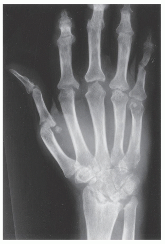
FIGURE 15.12 Mixed connective tissue disease. A 44-year-old woman presented with clinical and imaging features of rheumatoid arthritis. In addition, she had clinically documented dermatomyositis. A dorsovolar radiograph of her left hand shows extensive articular erosions at radiocarpal, metacarpophalangeal, and proximal interphalangeal joints, typical for rheumatoid arthritis. The muscle biopsy result was consistent with polymyositis.


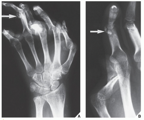
FIGURE 15.13 Mixed connective tissue disease. A 26-year-old woman presented with swelling of both hands, polyarthralgia, and Raynaud phenomenon. She tested positively for the rheumatoid factors and ANAs, and her clinical findings were characteristic for SLE and scleroderma. Oblique radiograph (A) of the right hand and coned-down view (B) of the thumb and index finger of the left hand show flexion deformities and subluxations in the multiple joints. Deformities of both thumbs are characteristic for SLE, whereas soft-tissue calcifications (arrows) are typical for scleroderma. The clinical diagnosis was MCTD.


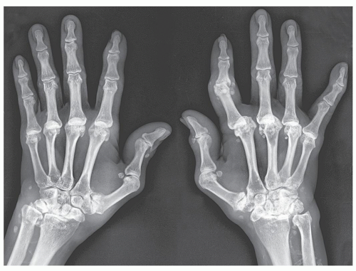
FIGURE 15.14 Mixed connective tissue disease. Dorsovolar radiograph of the hands of a 55-year-old woman with documented long-standing rheumatoid arthritis, lupus erythematosus, and scleroderma shows erosive changes in both wrists, subluxations in the metacarpophalangeal joints, and soft-tissue calcifications.


TABLE 15.2 Clinical and Imaging Hallmarks of Metabolic, Endocrine, and Miscellaneous Arthritides


Type of Arthritis


Site


Crucial Abnormalities


Technique/Projection


Gout (M > F)


Great toe


Large joints (knee, elbow)


Hand


Articular erosion with preservation of part of joint


Overhanging edge of erosion


Lack of osteoporosis


Periarticular swelling


Standard views of affected joints


Tophi


Dual-energy color-coded CT


CPPD crystal deposition disease (M = F)


Variable joints


Chondrocalcinosis (calcification of articular cartilage and menisci)


Calcifications of tendons, ligaments, and capsule


Standard views of affected joints


Femoropatellar joint


Joint space narrowing


Subchondral sclerosis


Osteophytes


Lateral (knee) and axial (patella) views


Wrists, elbows, shoulders, ankles


Degenerative changes with chondrocalcinosis


Standard views of affected joints


CHA crystal deposition disease (F > M)


Variable joints, but predilection for shoulder joint (supraspinatus tendon)


Pericapsular calcifications


Calcifications of tendons


Standard views of affected joints


Hemochromatosis (M > F)


Hands


Involvement of second and third metacarpophalangeal joints with beak-like osteophytes


Dorsovolar view


Large joints


Chondrocalcinosis


Standard views of affected joints


Alkaptonuria (ochronosis) (M = F)


Intervertebral disks, sacroiliac joints, symphysis pubis, large joints (knees, hips)


Calcification and ossification of intervertebral disks, narrowing of disks, osteoporosis, joint space narrowing, periarticular sclerosis


Anteroposterior and lateral views of spine; standard views of affected joints


Hyperparathyroidism (F > M)


Hands


Destructive changes in interphalangeal joints


Subperiosteal resorption


Dorsovolar view


Dorsovolar and oblique views


Multiple bones


Skull


Spine


Bone cysts (brown tumors)


Salt-and-pepper appearance


Rugger-jersey appearance


Standard views specific for locations


Lateral view


Lateral view


Acromegaly (M > F)


Hands


Widened joint spaces


Large sesamoid


Degenerative changes (beak-like osteophytes)


Dorsovolar view


Skull


Facial bones


Heel


Spine


Large sinuses


Large mandible (prognathism)


Thick heel pad (>25 mm)


Thoracic kyphosis


Lateral view


Lateral view


Lateral view


Lateral view (thoracic spine)


Amyloidosis (M > F)


Large joints (hips, knees, shoulders, elbows)


Articular and periarticular erosions, osteoporosis (periarticular), joint subluxations, pathologic fractures


Standard views of affected joints


Radionuclide bone scan (scintigraphy)


Multicentric reticulohistiocytosis (F > M)


Hands (distal and proximal interphalangeal joints)


Feet


Soft-tissue swelling, articular erosions, lack of osteoporosis


Dorsovolar view


Norgaard (ball-catcher’s) view


Dorsoplantar view


Oblique view


Hemophilia (M > F)


Large joints (hips, knees, shoulders)


Elbows, ankles


Joint effusion, osteoporosis, symmetrical and concentric joint space narrowing, articular erosions, widening of intercondylar notch, squaring of patella; very similar to changes of juvenile rheumatoid arthritis


Standard views of affected joints


MRI


M, male; F, female; CT, computed tomography; CPPD, calcium pyrophosphate dihydrate; CHA, calcium hydroxyapatite; MRI, magnetic resonance imaging.


Hyperuricemia

An increased miscible pool of uric acid with resulting hyperuricemia can occur in two principal ways. First, urate is produced in such large quantities that, even though excretion routes are of normal capacity, they are inadequate to handle the excessive load. Second, the capacity for uric acid excretion is critically reduced so that even a normal quantity of uric acid cannot be eliminated.

In 25% to 30% of gouty patients, a primary defect in the rate of purine synthesis causes excessive uric acid formation, as reflected in excessive urinary uric acid excretion (more than 600 mg/day) measured while the patient is maintained on a standard purine-free diet. Increased production can also be seen in gout secondary to myeloproliferative disorders associated with increased destruction of cells and result in increased breakdown of nucleic acids. Decreased excretion occurs in primary gout in patients with a dysfunction in the renal tubular capacity to excrete urate and in patients with chronic renal disease. In most patients, however, there is evidence of both uric acid overproduction and diminished renal excretion of uric acid.


The chance of development of gouty arthritis in hyperuricemic individuals should increase in proportion to the duration and, even more, to the degree of hyperuricemia. Monosodium urate, however, has a marked tendency to form relatively stable supersaturated solutions; therefore, the proportion of hyperuricemic patients in whom gouty arthritis actually develops is relatively low. The clinical development of gouty arthritis in the hyperuricemic subject is also substantially influenced by other factors, such as binding of urate to plasma proteins or the presence of promoters or inhibitors of crystallization.


Examination of Synovial Fluid

A wet preparation of fresh synovial fluid is best for the examination of crystals. Although crystals may often be seen by ordinary light microscopy, reliable identification requires polarization equipment. To differentiate between urate and pyrophosphate crystals—characteristics of gout and pseudogout, respectively—a compensated, polarized light microscope is advisable. Because both types of crystals are birefringent, they refract the polarized light that passes through them. The birefringence phenomenon is caused by the refractive index for light, which vibrates either parallel or perpendicular to the axis of the crystal being viewed. Color is the key to negative or positive birefringence. Urates are strongly birefringent; therefore, they are brightly colored in polarized light, with a red compensator. They are usually seen as needles. During an acute gouty attack, many intra-leukocytic crystals are present. Monosodium urate crystals are negatively birefringent, that is, they appear yellow when the longitudinal axis of the crystal is parallel to the axis of slow vibrations of the red compensator on the polarizing system, and they appear blue when perpendicular.

Monosodium urate crystals, the pathogens of gouty arthritis, range in length from 2 to 10 µm and are found within synovial leukocytes or extracellularly in virtually every case of acute gout, although the likelihood of finding such crystals varies inversely with the amount of time elapsed from the onset of symptoms to the time of examination. Crystals from tophi may be larger.


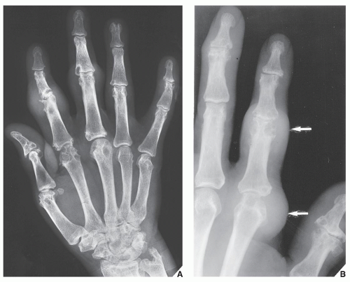
FIGURE 15.15 Gouty arthritis. (A) Dorsovolar radiograph of the left hand of a 43-year-old man with tophaceous gout shows multiple sharply marginated articular and periarticular erosions and soft-tissue masses at the proximal interphalangeal joints of the index and middle fingers, representing tophi. (B) Dorsovolar radiograph of the fingers of a 70-year-old man with gouty arthritis shows multiple articular and periarticular erosions associated with large tophi (arrows).


Imaging Features

Gouty arthritis has several characteristic imaging features. Erosions, which are usually sharply marginated, are initially periarticular in location and are later seen to extend into the joint (Fig. 15.15); an “overhanging edge” of erosion is a frequent identifying feature (Figs. 15.16 and 15.17). Occasionally, intraosseous defects are present secondary to the formation of intraosseous tophi (Figs. 15.18 and 15.19). Usually, there is a striking lack of osteoporosis, which helps differentiate this condition from rheumatoid arthritis. The reason for the absence of osteoporosis is that the duration of an acute gouty attack is too short to allow the development of the disuse osteoporosis so often seen in patients with rheumatoid arthritis. If erosion involves the articular end of the bone and extends into the joint, part of the joint is usually preserved (Fig. 15.20; see also Fig. 15.16). Unlike rheumatoid arthritis, periarticular and articular erosions are asymmetric in distribution (Fig. 15.21). In chronic tophaceous gout, sodium urate deposits in and around the joint are seen, creating a dense mass in the soft tissues called a tophus, which frequently exhibits calcifications (Figs. 15.22, 15.23, 15.24; see also Figs. 15.15 and 15.16). Characteristically, tophi are randomly distributed and are usually asymmetric; if they occur in the hands or feet, they are more often seen on the dorsal aspect (Fig. 15.25). Currently dual-energy color-coded CT images can accurately depict gouty tophi (Fig. 15.26 and 15.27; see also Figs. 2.15, 12.10, and 12.11). Reported sensitivity of this technique varies between 78% and 100% and specifically between 89% and 100%. Tophaceous gouty deposits exhibit low signal intensity on magnetic resonance imaging (MRI) in all pulse sequences and strong enhancement following intravenous injection of gadolinium (see Fig. 15.19C,D).

Only gold members can continue reading. Log In or Register to continue

Stay updated, free articles. Join our Telegram channel

Jul 24, 2016 | Posted by in MUSCULOSKELETAL IMAGING | Comments Off on Miscellaneous Arthritides and Arthropathies

Full access? Get Clinical Tree

Get Clinical Tree app for offline access