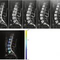
Fig. 3.1
Normal spinal bone marrow, 2 months.
Sagittal T1-weighted image (left) of the spine of a 2-month-old boy shows biconvex shape of vertebral bodies with large hyperintense cartilaginous endplates (square). Matching T2-weighted image (right) is shown for comparison. Also shown is tethered cord with spina bifida and lipocele (asterisk) (Courtesy of Ioannis Nikas, M.D., Children’s Hospital “Aghia Sophia,” Athens, Greece)
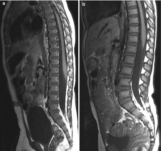
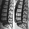
Fig. 3.2
Normal spinal bone marrow, 8 and 26 months.
Sagittal T1-weighted images of the spine of an 8-month-old boy (a) and a 26-month-old girl (b) show that the vertebral bodies are at least isointense to the intervertebral discs. Note the more square-shaped appearance of the vertebrae in b (Courtesy of Ioannis Nikas, M.D., Children’s Hospital “Aghia Sophia,” Athens, Greece)
Key Point
Spinal bone marrow is hypointense to intervertebral discs in the first few months of life
The gradual increase in the signal intensity of spinal bone marrow reflects the increase in fat content, a process that continues throughout life. The distribution of yellow marrow in the spine may not, however, be homogeneous; four different patterns of yellow marrow deposition in the spine have been described based on MR images [3]:
Pattern 1: Fatty marrow of variable thickness is seen surrounding the basivertebral vein and parallel to it (Fig. 3.3).
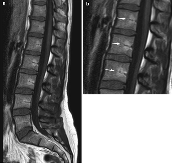
Fig. 3.3
Spinal red to yellow marrow conversion, pattern 1 (basivertebral).
Sagittal T1-weighted image (a) and detailed view (b) of the lumbosacral spine of a 25-year-old woman show hyperintense fatty marrow (arrows) around basivertebral vessels
Pattern 2: Fatty marrow appears along the vertebral endplates. This pattern is more frequently seen in the lumbar and cervical spine, which provide major support to the body and are more often subjected to mechanical stress resulting in degenerative changes (Modic II endplate changes) than the thoracic spine which is assisted by the rib cage (Fig. 3.4).
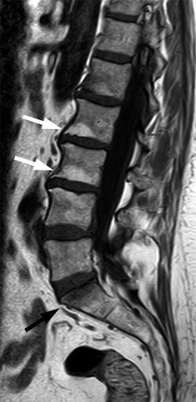
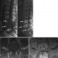
Fig. 3.4
Spinal red to yellow marrow conversion, pattern 2 (endplate).
Sagittal T1-weighted image of the lumbosacral spine of a 59-year-old woman shows hyperintense fatty marrow along the inferior endplates of L2 and L3 (Modic II degenerative endplate changes, white arrows). Also note Modic I degenerative change along the superior endplate of S1 (black arrow)
Pattern 3: Rounded or oval-shaped foci of fat are seen throughout the spine. In this pattern, distinct fatty marrow foci intermingle with red marrow giving the spine a mosaic-like appearance (Fig. 3.5). Foci of fat may become more confluent, alternating with smaller ill- or well-defined rests of red marrow (Fig. 3.6).
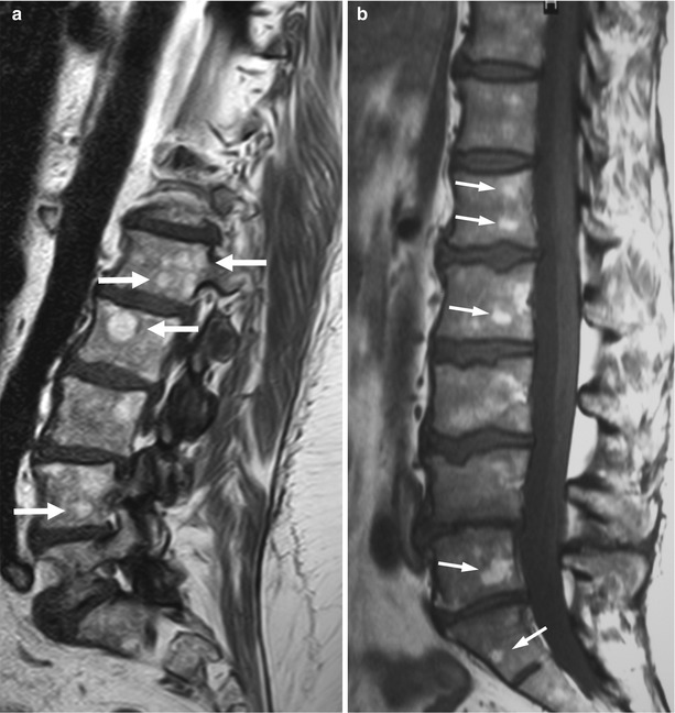
Fig. 3.5
Spinal red to yellow marrow conversion, pattern 3 (mosaic, distinct).
Sagittal T1-weighted images of the lumbosacral spine of a 58-year-old woman (a) and a 48-year-old woman (b) show multiple, well-defined, hyperintense foci of yellow marrow (arrows)
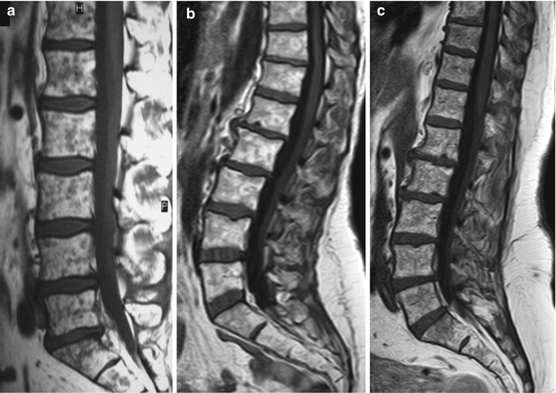
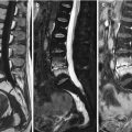
Fig. 3.6
Spinal red to yellow marrow conversion, pattern 3 (mosaic, confluent).
Sagittal T1-weighted images of the lumbosacral spine in three different individuals show confluent areas of fatty marrow with residual, small, well-defined (a) or ill-defined (b, c) hypointense red marrow foci
Pattern 4: Homogeneous fatty spine (Fig. 3.7). This pattern is most helpful when searching for bone marrow lesions, since it facilitates their detection as hypointense foci against a “bright” background.
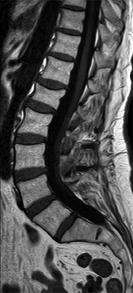
Fig. 3.7
Spinal red to yellow marrow conversion, pattern 4 (homogeneous).
Sagittal T1-weighted image of the lumbosacral spine of a 75-year-old woman shows homogeneous high signal intensity of fatty marrow
In some cases, there may be a combination of patterns, most often 2 and 3. Pattern 4 is more frequently observed in older individuals.
Key Point
In the spine, four patterns of fatty marrow deposition are described: basivertebral, endplate, mosaic, and homogeneous
3.2 Pelvis
The pelvis, as part of the central skeleton, is a site of persistent red marrow in the adult. Ricci et al. described islands of fatty marrow first appearing at the superomedial aspect of the acetabulum and later in the iliac bones and around the sacroiliac joints [3]. Marrow heterogeneity of the acetabulum and anterior iliac bone with increased deposition of fatty marrow is common in adolescents [4]. In the sacrum, presence of fatty marrow is more pronounced in the periphery of the sacral wings, presumably because of the peripheral to central pattern of red to yellow marrow conversion in the skeleton [5] (Fig. 3.8).
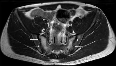
Fig. 3.8


Red to yellow marrow conversion, sacrum.
Related posts:

Stay updated, free articles. Join our Telegram channel
Full access? Get Clinical Tree
