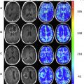
Magnetic resonance spectroscopy (MRS) is a magnetic resonance–based imaging modality that allows noninvasive sampling of metabolic changes in normal and abnormal brain parenchyma. MRS is particularly useful in the differentiation of developmental or non-neoplastic disorders from neoplastic processes. MRS is also useful during routine imaging follow-up after radiation treatment or during antiangiogenic treatment and for predicting outcomes and treatment response. The objective of this article is to provide a concise but thorough review of the basic physical principles, important applications of MRS in brain tumor imaging, and future directions.
Key points
- •
Magnetic resonance spectroscopy (MRS) is a noninvasive technique that allows the study of metabolic processes and chemical environment in the brain parenchyma.
- •
MRS is one of the few diagnostic techniques that can be used for evaluation of low-grade neoplastic processes and for their differentiation from non-neoplastic entities.
- •
Despite many technical and reimbursement challenges to its use in routine clinical practice, MRS will continue to develop as an important and sensitive imaging tool for assessment of intracranial pathologies.
Discussion of problem/clinical presentation
MRS allows the qualitative and quantitative assessment of specific metabolites in the brain parenchyma or intracranial extra-axial spaces. MRS analysis of brain tumors can be performed using 1 H (proton) MRS or, less frequently, with 31 P (phosphorus) or 13 C (carbon) MRS techniques. For 1 H MRS, the most common metabolites evaluated in routine clinical practice include N -acetyl aspartate (NAA), choline-containing compounds (Cho), creatine (Cr), myo-inositol (mI), lipid (Lip), and lactate (Lac) ( Table 1 ). NAA is considered a neuronal metabolite and is decreased in processes with neuronal destruction or dysfunction. Cr is a metabolite related to the cellular energy metabolism and is considered relatively stable in different pathologic processes affecting the central nervous system and useful as a reference metabolite. Cho are related to membrane turnover and their elevation is indicative of a process that results in increased glial proliferation and membrane synthesis (as seen with cellular proliferative disorders). Lip peaks are often indicative of areas of necrosis and Lac peaks are directly originated from processes resulting in anaerobic metabolism. mI can be a marker of astrocytic metabolism and can be seen elevated in certain pathologic processes (see Table 1 ).
| Metabolite | Peak Configuration | Resonance (ppm) | Best Echo Time for Detection | Clinical Associations |
|---|---|---|---|---|
| NAA | Singlet | 2.0 | Short or long TE | Neuronal marker (not seen in non-neural brain tumors) |
| Cho | Singlet | 3.22 | Short or long TE | Membrane turnover marker and cellular proliferation |
| Cr | Singlets | 3.03 and 3.9 | Short or long TE | Cellular energy byproduct. Lower in necrosis |
| mI | Multiplets | 3.56 | Short TE | Low-grade gliomas, gliomatosis |
| Lip | Broad peaks | 0.9, 1.3 | Short TE | Tuberculomas, PCNSL, radiation necrosis |
| Lac | Doublet | 1.33 | 1.5T = inverted at 135–144 ms 3T = 288 ms | Anaerobic metabolism marker Prominent if necrosis or hypoxia |
| Glx | Multiplets | 2.1–2.4 ppm; 3.7 ppm | Short TE | Detected in GBM, astrocytomas and oligodendrogliomas |
| Taurine | Triplets | 3.4 | Short TE | Medulloblastomas |
| Alanine | Doublet | 1.47 | 1.5 T = 144 ms | Meningiomas |
| Citrate | Multiplets | 2.6 | 3T = 35 ms and inverts at 97 ms | Gliomas, particularly aggressive pediatric types |
| Gly | Singlet | 3.55 | 3T = 160 ms | Low-grade gliomas, central neurocytomas |
| 2HG | Multiplets | 1.85, 2.01, 2.28, and 4.05 | Best seen with spectral editing techniques 3T = 97 ms | IDH mutations |
Common diagnostic problems encountered in routine clinical brain tumor imaging can be summarized as follows.
Is It Neoplastic or Not?
Non-neoplastic processes, including malformations of cortical development (such as focal cortical dysplasia) ( Fig. 1 ), hamartomas, cerebral infarcts, infectious pathologies, inflammatory diseases (including demyelinating and vasculitic processes), and vascular pathologies (including capillary telangiectasias and cavernous malformations), can be difficult to differentiate from intra-axial or extra-axial intracranial neoplastic processes in conventional magnetic resonance (MR) studies ( Table 2 ). MRS is a useful imaging tool to help in the differentiation and characterization of these pathologies. Neoplastic processes have metabolic byproducts related to their mitotic activity (Cho) and neuronal dysfunction (NAA) that can be detected by MRS and improve the accuracy of the clinical diagnosis ( Fig. 2 ). The closer the MR spectrum is to a normal spectrum the more likely that the intracranial lesion is a benign process or developmental anomaly (see Fig. 1 ). There is significant overlap in the Cho/NAA ratios, however, between non-neoplastic processes, such as tumefactive demyelinating lesions, infarcts, and infectious processes with neoplastic pathologies. Specific metabolic markers have been identified that may make this distinction more reliable (eg, glutamate/glutamine [Glx] for demyelination or 2-hydroxyglutarate [2HG] for isocitrate dehydrogenase [IDH] 1–mutant gliomas).

| Pathology | N -Acetyl Aspartate | Choline | Myo-inositol | Glutamate/Glutamine | Glycine | Lactate | Lipid | Taurine | Alanine | Amino Acids | Succinate | 2-Hydroxyglutarate | Comments |
|---|---|---|---|---|---|---|---|---|---|---|---|---|---|
| Glioblastoma and anaplastic gliomas | −−/−−− | ++/+++ | ++ | ++ | + | Necrotic areas with high Lac and Lip | |||||||
| Diffuse astrocytoma | − | + | ++ | + | + | ||||||||
| Pilocytic astrocytoma | − | + | + | + | Decreased Cr | ||||||||
| Medulloblastoma | − | ++/+++ | + | +/++ | + | + | +/++ | Groups 3 and 4 show high taurine, lower Lip, and high Cr. SHH tumors show high Cho and Lip, with minimal taurine. Also phosphocholine | |||||
| Ependymoma | ++/+++ | + | Also glycerophosphocholine in vitro | ||||||||||
| Intracranial metastases | −− | ++ | ++ | Relatively normal metabolites in the parenchyma surrounding the lesion Absent NAA within the lesion | |||||||||
| PCNSL | +++ | MRS obtained from non-necrotic lesions | |||||||||||
| Epidermoid cysts | ++ | + | + | Aminoacids: valine, isoleucine and glycine | |||||||||
| Meningioma | −−/−−− | ++ | + | Alanine (up to 90% of cases) | |||||||||
| Piogenic abscess | −/−− | +/++ | ++ | ++ | ++ | Aminoacids, Lac, alanine, and acetate | |||||||
| Tuberculoma | − | + | ++ | + | ++ | Higher Cho/Cr and mI/Cr in tumors. Also peak at 3.8 ppm, possibly guanidinoacetate | |||||||
| Tumefactive demyelinating lesion (TDL) | −/−− | +/++ | + | ||||||||||
| Radiation necrosis | − | + | ++ | ++ | Higher Cho/Cr and mI/Cr in tumors |

Is the Lesion a Primary or a Secondary Brain Tumor?
Several studies have shown the utility of MRS, particularly using the multivoxel technique for differentiation of glioblastoma from an intracerebral metastasis. The assessment of normal-appearing brain parenchyma in the immediate vicinity of the tumor has been shown reliable, with glioblastoma cases showing higher Cho/NAA ratios compared with metastatic lesions ( Figs. 3 and 4 ).


Is It a High-Grade or Low-Grade Tumor?
Multiple studies have shown the value of MRS for predicting the histologic grade of glial tumors. Higher Cho/NAA ratios have been associated with higher World Health Organization (WHO) grades among glial tumors (see Figs. 2 and 3 ). The Cho/Cr ratio seems more accurate for the differentiation of high-grade versus low-grade gliomas (with sensitivity and specificity values of 80% and 76%, respectively).
Where Is the Best Target for Surgery or Treatment?
MRS can provide a general overview of the metabolic profile of a brain tumor and indicate areas of higher clinical aggressiveness or histologic grade (with higher Cho/Cr ratios). Multivoxel MRS can guide the neurosurgeon for sampling the anatomic sites with the highest histologic grade in low-grade tumors and increases the diagnostic accuracy of stereotactic and excisional biopsies (see Fig. 3 ). There is also growing evidence of the potential use of 3-D volumetric MRS imaging (MRSI) to guide radiation treatment. MRS can potentially identify areas with abnormal Cho/NAA ratios that can extend beyond the tumoral margins defined by conventional MR sequences (see Fig. 3 ).
Radiation-Induced Changes or Recurrent High-Grade Tumor?
MRS, particularly when using multivoxel techniques, has been shown useful for the differentiation of radiation necrosis and recurrent high-grade glioma and recommended as an evidence level II diagnostic modality for this differentiation. In general, areas with recurrent glioma have higher Cho/NAA ratios with variable Lip peaks. Pure radiation necrosis shows prominent Lip with relative decrease of the remaining metabolites compared with the contralateral normal appearing brain parenchyma ( Fig. 5 ). Relative ratios of intralesional Cho to contralateral Cr have also been shown to be a good marker to distinguish between radiation necrosis and recurrent high-grade tumor. This distinction is not always black and white, however, because many cases have concurrent areas of neoplastic involvement and postradiation changes. In addition, elevated Lip can also be seen in high-grade gliomas near areas of central necrosis.
How to Identify Infiltrative (Recurrent) Tumor in Patients with Glioblastoma Treated with Antiangiogenic Treatment
Patients receiving antiangiogenic medications, such as bevacizumab, show rapid decrease or resolution of the intratumoral abnormal enhancement and the conventional MR sequences are difficult to interpret during the follow-up imaging of these cases. MRS has the potential to identify abnormal metabolic patterns suggestive of tumoral infiltration in progressive areas of fluid-attenuated inversion recovery (FLAIR) hyperintensity in patients treated with bevacizumab, despite the lack of abnormal enhancement. A multicenter trial by the American College of Radiology Imaging Network demonstrated increased NAA/Cho and decreased Cho/Cr levels 8 weeks into the treatment with bevacizumab in patients with recurrent glioblastoma correlated with increased progression free survival rates.
Can Treatment Response or Outcomes Be Predicted with Magnetic Resonance Spectroscopy?
Multiple studies have highlighted the potential role of MRS to predict treatment responses and outcome after treatment. The identification of 2HG in glial tumors with IDH1 mutations and glutamate in pediatric medulloblastomas is associated with better overall survival rates. The decrease of 2HG within glial tumors after treatment also correlate with functional status. MRS, specifically the Lac-to-NAA ratio, may also be helpful in the prediction of potential sites of glioblastoma recurrence. Increased Cho/NAA values 3 weeks after radiation treatment are associated with higher probability of early glioblastoma progression.
Discussion of problem/clinical presentation
MRS allows the qualitative and quantitative assessment of specific metabolites in the brain parenchyma or intracranial extra-axial spaces. MRS analysis of brain tumors can be performed using 1 H (proton) MRS or, less frequently, with 31 P (phosphorus) or 13 C (carbon) MRS techniques. For 1 H MRS, the most common metabolites evaluated in routine clinical practice include N -acetyl aspartate (NAA), choline-containing compounds (Cho), creatine (Cr), myo-inositol (mI), lipid (Lip), and lactate (Lac) ( Table 1 ). NAA is considered a neuronal metabolite and is decreased in processes with neuronal destruction or dysfunction. Cr is a metabolite related to the cellular energy metabolism and is considered relatively stable in different pathologic processes affecting the central nervous system and useful as a reference metabolite. Cho are related to membrane turnover and their elevation is indicative of a process that results in increased glial proliferation and membrane synthesis (as seen with cellular proliferative disorders). Lip peaks are often indicative of areas of necrosis and Lac peaks are directly originated from processes resulting in anaerobic metabolism. mI can be a marker of astrocytic metabolism and can be seen elevated in certain pathologic processes (see Table 1 ).
| Metabolite | Peak Configuration | Resonance (ppm) | Best Echo Time for Detection | Clinical Associations |
|---|---|---|---|---|
| NAA | Singlet | 2.0 | Short or long TE | Neuronal marker (not seen in non-neural brain tumors) |
| Cho | Singlet | 3.22 | Short or long TE | Membrane turnover marker and cellular proliferation |
| Cr | Singlets | 3.03 and 3.9 | Short or long TE | Cellular energy byproduct. Lower in necrosis |
| mI | Multiplets | 3.56 | Short TE | Low-grade gliomas, gliomatosis |
| Lip | Broad peaks | 0.9, 1.3 | Short TE | Tuberculomas, PCNSL, radiation necrosis |
| Lac | Doublet | 1.33 | 1.5T = inverted at 135–144 ms 3T = 288 ms | Anaerobic metabolism marker Prominent if necrosis or hypoxia |
| Glx | Multiplets | 2.1–2.4 ppm; 3.7 ppm | Short TE | Detected in GBM, astrocytomas and oligodendrogliomas |
| Taurine | Triplets | 3.4 | Short TE | Medulloblastomas |
| Alanine | Doublet | 1.47 | 1.5 T = 144 ms | Meningiomas |
| Citrate | Multiplets | 2.6 | 3T = 35 ms and inverts at 97 ms | Gliomas, particularly aggressive pediatric types |
| Gly | Singlet | 3.55 | 3T = 160 ms | Low-grade gliomas, central neurocytomas |
| 2HG | Multiplets | 1.85, 2.01, 2.28, and 4.05 | Best seen with spectral editing techniques 3T = 97 ms | IDH mutations |
Stay updated, free articles. Join our Telegram channel

Full access? Get Clinical Tree


