, Carlos Capuñay1, Carlos E. Sueldo2 and Juan Mariano Baronio3
(1)
Diagnóstico Maipú, Buenos Aires, Argentina
(2)
University of California, San Francisco, CA, USA
(3)
CEGYR, Buenos Aires, Argentina
In the 6th week of the embryo development the morphological sexual characteristics of both sexes start to establish themselves. In every embryo, on both sides of the middle line, the following structures are present: (i) a genital septum; (ii) a Wolf mesenteric duct, beside the genital septum; (iii) a paramesonephric Müllerian duct that consists of three segments: (a) a vertical cranial sector beside the Wolf duct; (b) a middle horizontal sector that passes in front of the Wolf duct; and (c) an inferior or caudal sector. According to the established sex at the moment of fertilization, the absence of the Y chromosome clears the way for the differentiation in the female sense, establishing the retraction of the Wolf ducts, the development of the structures derived of the Müller paramesonephric ducts and the formation of the ovaries [1–3].
The uterovaginal primordial is formed from the fusion of the Müllerian ducts. They have a Y shape, with a medial and caudal direction reaching the urogenital middle. The cranial and middle sectors are the origins of the Fallopian tubes, while the caudal portion forms the uterus and 4/5 of the vagina. The fusion begins in the inferior section with a caudal-cephalic direction, completing itself in 11th or 13th week of the mentioned period. Once finalized and the uterine silhouette formed, the reabsorption of the septum which divides both conduits to constitute the endometrial cavity, the cervical canal and part of the vagina begins (Fig. 4.1). This phase concludes after the 3rd month of the fertilization [2, 4–6].
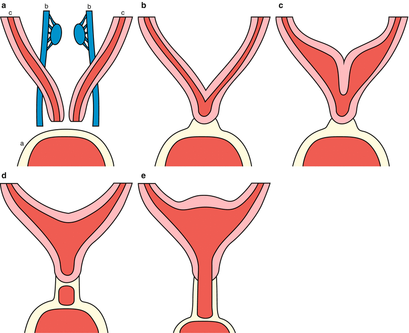

Fig. 4.1
Embryologic development. (a–e) Process of female sexual differentiation (a urogenital septum; b Wolf mesonferic duct; c paramesonephric Müllerian duct)
In this chapter, the normal feminine reproductive apparatus and its radiological manifestation in virtual hysterosalpingography (VHSG) studies is described.
Uterus
The uterus is a muscular organ, of triangular shape located in the lower pelvis behind the bladder and in front of the rectum, with the cranial base and the neck on its inferior side, extending to the vagina. It possesses a virtual cavity, and its size varies depending on the age of the patient and her gestational history. In the nulliparous women it measures approximately 9 × 6 × 3 cm. It is formed of three layers: (i) the serose one, referring to the visceral leaf of the peritoneum that covers it; (ii) the myometrium, which conforms the middle layer and is characterized by the presence of intertwined smooth muscular fibers; (iii) the endometrium, which constitutes the inner glandular mucosa layer. The uterus divides into: cervix, body or corpus, and the isthmus as barrier between the first two segments.
Position of the Uterus
The position of the uterus is determined by the orientation of the neck in relation to the vagina (version) and the body in relation to the neck (flexion) [7]. The most common, anteversio-anteflexio, occurs when the neck is flexed forward over the vagina, and in the same way, the uterine body over the neck. The uterus can adopt different positions considered as normal variants. These are shown in Fig. 4.2
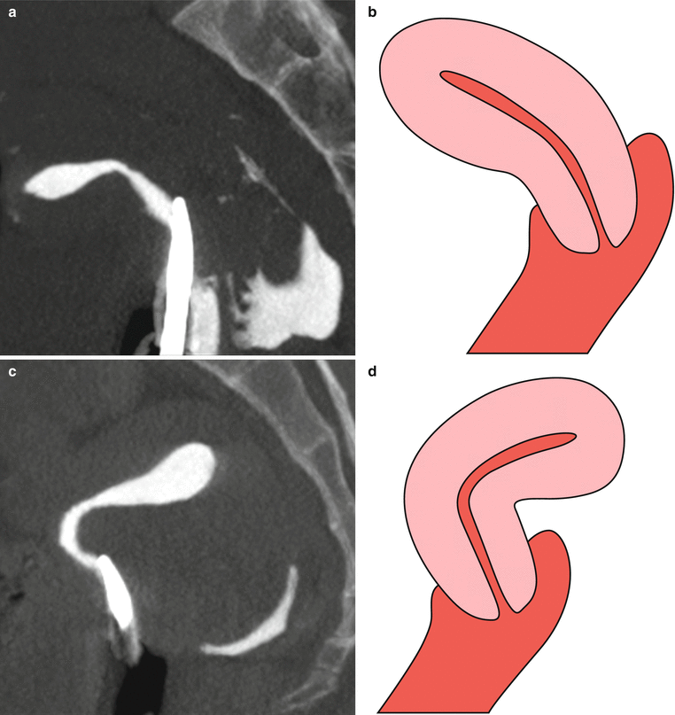
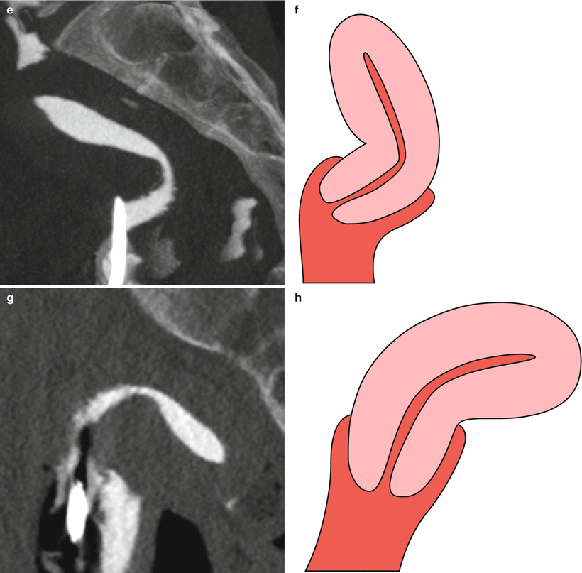


Fig. 4.2
Position of uterus in the lower pelvis. Maximum intensity projection VHSG images which show the different positions, determined by the orientation of the cervical neck in relation to the vagina and of the body in relation to the cervical neck. (a, b) Anteversio-anteflexio uterus. (c, d) Anteversio-retroflexio uterus. (e, f) Retroversio-anteflexio uterus. (g, h) Retroversio-retroflexio uterus
Cervix and Isthmus
The uterine cervix is the lowest fibromuscular region of the uterus, thin and round with an average length of 4 cm and a diameter of 2.5 cm. Its lower part denominated vaginal portion or tenca snout, penetrates in the vagina while the upper portion remains on top and joins the muscular body of the uterus at the level of the internal cervical orifice in the isthmic region. The shape and size of the uterine neck suffers variations according to the age, moment of menstrual cycle and number of pregnancies of the patient. In a nulliparous women, the neck is fusiform and the external os is small, round and located in the center; on the other hand, in a woman who has already had a child, it is of great volume and the external os presents the aspect of a wide elongated clift.
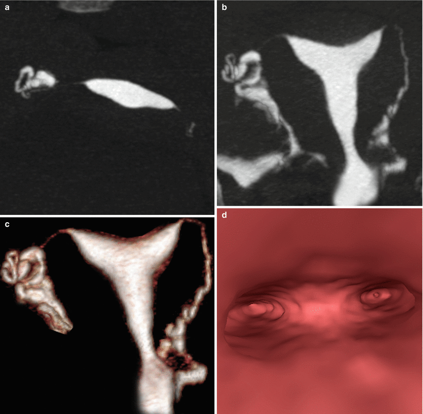
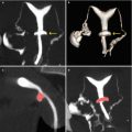
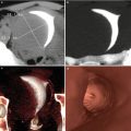
The cervical canal passes through the endocervix. It communicates the vagina with the uterine cavity through the internal and external cervical os. Its length and diameter present a great variation according to the age and the moment of the patient’s hormonal cycle (Fig. 4.3). The cervical canal is covered by the cylindrical epithelium, also known as the glandular epithelium. This epithelium is not a smooth and flat surface in the canal, it is made of multiplane longitudinal folds that project towards the lumen of the canal and give place to papillary projections. At the same time, it produces in the cervical estroma invaginations that constitute crypts, also known as endocervical glands (Fig. 4.4). In the VHSG studies, these glands are seen as sacular structures of small size projected on the walls of the cervical canal. On occasions, when dilated, they can be prominent, and observed as pseudodiverticular outgrowths (Fig. 4.5).
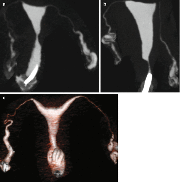
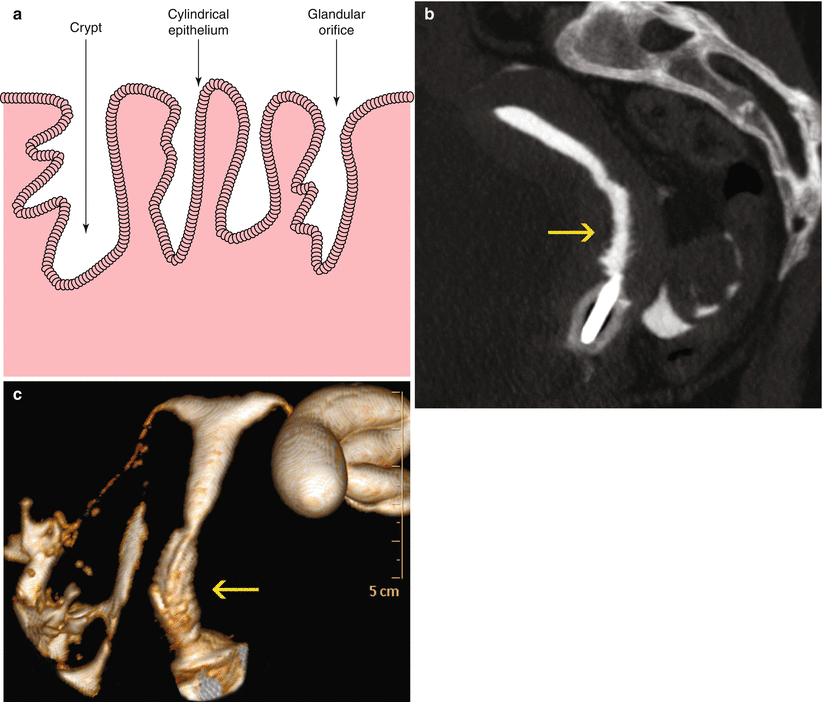
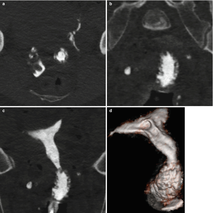
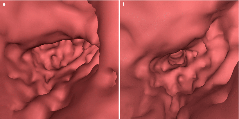

Fig. 4.3
VHSG study. Uterine cervical neck and normal variants. (a) Coronal maximum intensity projection (MIP) image which shows a cervical canal of normal caliber and morphology. (b) Coronal MIP image which shows a narrow cervical canal. (c) 3D volume rendering image which shows a wide cervical canal with normal fold visualization

Fig. 4.4
Epithelium lining of the cervical canal. (a) Display which shows the epithelial lining of the cervical canal, with longitudinal folds which project towards the lumen and give origin to papillary projections and invaginations or crypts. (b, c) Sagittal maximum intensity projection and coronal 3D volume rendering images which show the characteristic longitudinal folds of the cervical canal (arrows)


Fig. 4.5
VHSG study. Large cervical canal with prominent glands. (a) Axial CT image. (b) Coronal multiplanar reconstruction image. (c) Coronal maximum intensity projection image. (d) Coronal oblique 3D volume rendering image. (e, f) Virtual endoscopy images
The isthmus is the shortest portion of the uterus, located between the upper part of the neck and the lower part of the uterine body (Fig. 4.6).
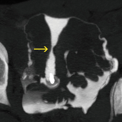

Fig. 4.6
Uterine isthmus (arrow). Coronal maximum intensity projection image
Body and Fundus
The uterine fundus conforms the upper dome-like portion of the uterus. It is constituted by a thick layer of muscular fibers, and is located on top of the insertion zone of the Fallopian tubes. The external configuration of the uterus is of utmost importance in the classification of uterine malformations. The final result of the complete fusion of the müllerian ducts during the embrionary period determines the external convex configuration. The uterine body corresponds to the principal portion of the uterus, containing a unique endometrial cavity, of triangular shape and covered by the endometrium (Fig. 4.7). The alterations in the process of fusion of the paramesonferic ducts that determine the appearance of the external uterine fundus, plus the alterations that occur during the process of reabsorption of the septum that divides the same and that lead to changes in the cavity, give way to the different uterine malformations [3–6, 8, 9]. See Chap. 8.

