Orientation to Scanning


Orientation to Scanning


Diane M. Kawamura


This chapter focuses on the sonography examination of the abdomen and superficial structures. It was written to assist sonographers in acquiring, using, and understanding the sonographic imaging terminology used in the remainder of this textbook. Accurate and precise terminology allows effective communication among professionals.

The textbook is divided into five sections: introduction to sonography, abdominal sonography, superficial structure sonography, neonatal and pediatric sonography, and special study sonography.


ANATOMIC DEFINITIONS

The profession adopted standard nomenclature from the anatomists’ terminology to communicate anatomic direction. Table 2-1 and Figure 2-1 illustrate how these simple terms help avoid confusion and provide specific information. A person in the conventional anatomic position stands erect, with feet together, the arms by the sides, and the palms and face directed forward, facing the observer. When sonographers use directional terms or describe regions or anatomic planes, it is assumed that the body is in the anatomic position.

There are three standard anatomic planes (sections), which are imaginary flat surfaces passing through a body in the standard anatomic position. The sagittal plane and coronal plane follow the long axis of the body and the transverse plane follows the short axis of the body1 (Fig. 2-2).

The word sagittal literally means “flight of an arrow” and refers to the plane that courses vertically through the body and separates it into right and left portions. The plane that divides the body into equal right and left halves is referred to as the median sagittal or midsagittal plane. Any vertical plane on either side of the midsagittal plane is a parasagittal plane (para means “alongside of”). In most sonography cases, the term sagittal usually implies a parasagittal plane unless the term is specified as median sagittal or midsagittal. The coronal plane courses vertically through the body from right to left or left to right, and it divides the body into anterior and posterior portions. The transverse plane passes through the body from anterior to posterior and divides the body into superior and inferior portions and courses parallel to the surface of the ground.


SCANNING DEFINITIONS


Patient Position

Positional terms refer to the patient’s position relative to the surrounding space. For sonographic examinations, the patient position is described relative to the scanning table or bed (Table 2-2 and Fig. 2-3). In clinical practice, patients are scanned in a recumbent, semierect (reverse Trendelenburg or Fowler), or sitting position. On occasion, patients may be placed in other positions, such as the Trendelenburg (head lowered) or standing position, to obtain unobscured images of the area of interest. Sonographers frequently convey information on patient position and transducer placement simultaneously. This terminology most likely was adopted from radiography, where it describes the path of the X-ray beam through the patient’s body (projection), which results in a radiographic image (view). There is no evidence in the literature that this nomenclature has been adopted as a professional standard for sonographic imaging. Describing sonograms using the terms projection or view should be avoided. It is more accurate to describe the sonography image by stating the anatomic plane visualized, owing to the

transducer’s orientation (i.e., transverse). A more specific description of the image would include both the anatomic plane and the patient position (i.e., transverse, oblique).




Transducer Orientation

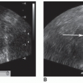
The transducer’s orientation as viewed on the monitor is the path of the insonating sound beam and the path of the returning echoes. Transducers are manufactured with an indicator (notch, groove, light) that is displayed on the monitor as a dot, arrow, letter of the manufacturer’s insignia, or other delineation. Scanning plane is the term used to describe the transducer’s orientation to the anatomic plane or to a specific organ or structure. The sonographic image is a representation of sectional anatomy. The term plane combined with the adjectives sagittal, parasagittal, coronal, and transverse describes the section of anatomy represented on the image (e.g., transverse plane).

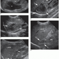
Because many organs and structures lie oblique to the imaginary body surface planes, sonographers must identify sectional anatomy accurately to utilize a specific organ and structure orientation for scanning surfaces. The sonography imaging equipment provides a lot of flexibility to rock, slide, and angle the transducer to obtain sectional images of organs oriented obliquely in the body. For example, to obtain the long axis of an organ, such as the kidney, the transducer is placed obliquely and is angled off of the standard anatomic positions: sagittal, parasagittal, coronal, or transverse plane. Sonographers frequently use the terms sagittal or parasagittal to mean longitudinal in depicting the anatomy in a long-axis section. Although some images in this text are labeled sagittal or parasagittal, they are, in fact, longitudinal planes because the image is organ specific. For
organ imaging, transverse planes are perpendicular to the long axis of the organ, and longitudinal and coronal planes are referenced to a surface. All three planes are based on the patient position and the scanning surface (Fig. 2-4A-C).


Image Presentation

When describing image presentation on the display monitor, the body, organ, or structure plane terminology, coupled with transducer placement, provides a very descriptive portrayal of the sectional anatomy being depicted. Current flexible, freehand scanning techniques may lack automatic labeling of the scanning plane. With the freehand scanning technique, quantitative labeling may be limited, which means reduced image reproducibility from one sonographer to another. Sonographers can usually select from a wide array of protocols for image annotation or employ postprocessing annotation. This is extremely important when the image of an isolated area does not provide other anatomic structures for a reference location. To ensure consistent practice, sonographers must correctly label all sonograms. With today’s equipment, standard presentation and labeling are easily achieved along with additional labeling of specific structures and added comments.

Only gold members can continue reading. Log In or Register to continue

Stay updated, free articles. Join our Telegram channel

Dec 10, 2022 | Posted by in ULTRASONOGRAPHY | Comments Off on Orientation to Scanning

Full access? Get Clinical Tree

Get Clinical Tree app for offline access