Orientation


Key Points


  • Providers must understand the orientation between patient, transducer, and ultrasound screen because ultrasound imaging generates two-dimensional images of three-dimensional structures.


  • Sagittal and coronal planes are along the long axis of the body, and these planes are often referred to as the longitudinal plane . The transverse plane is along the short axis of the body.


  • Providers can use real-time ultrasound to guide invasive procedures by tracking the needle tip using either a longitudinal (in-plane) or a transverse (out-of-plane) approach.


Introduction


Point-of-care ultrasound allows providers to perform focused exams at the bedside to answer specific clinical questions, guide management, and ultimately improve care of patients. This chapter focuses on orientation of providers to the ultrasound screen, transducer, and patient, as well as standard imaging planes. An important principle in bedside ultrasound imaging is understanding orientation of three-dimensional structures that are being displayed in two dimensions. A major advantage of real-time ultrasound scanning is the capability to visualize an object in multiple planes to better understand its three-dimensional structure.


Operator Orientation


Performing point-of-care ultrasound begins with operator orientation. Providers must use a systematic approach to consistently acquire images of high quality. Traditionally, providers have performed bedside scans standing on the left side of the bed, similar to the physical exam, with the ultrasound machine directly in front of them. One hand holds the transducer on the patient and the other hand operates the ultrasound machine. Providers might stand on the right side of the bed when scanning the heart due to the heart’s position in the left chest. The height of the patient’s bed and position of the ultrasound machine should be adjusted to optimize patient and operator comfort. The machine should be close to the bedside so that controls can be reached. The transducer should be held with the same hand, whether left or right, to help develop consistent habits and muscle memory.


Screen Orientation


During the early evolution of diagnostic ultrasound from the 1940s until the 1970s, general medical and cardiac ultrasound imaging developed two independent conventions for displaying images on the screen. In both conventions, the top of the screen corresponds with the probe face. Superficial structures are viewed at the top of the image and deeper structures are viewed at the bottom. The structure of interest should be maintained in the center of the screen for best image resolution with most portable ultrasound machines.


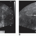
General medical ultrasound utilizes a convention with the transducer marker corresponding to the left side of the screen, usually depicted by a small colored circle or square on the screen ( Figure 4.1 ). The majority of specialties performing diagnostic ultrasound imaging follow this convention, including radiology and emergency medicine. Thus, imaging in a transverse plane generates cross-sectional images as if the patient were viewed from the foot of the bed; i.e., the liver is on the left, the spleen on the right. Some providers may choose to invert images generated by transvaginal ultrasound so that structures nearest the transducer are viewed at the bottom of the ultrasound screen.


Figure 4.1


Ultrasound image using conventional orientation with marker at left side of screen (arrow).


Cardiac ultrasound, on the other hand, utilizes a convention with the transducer marker corresponding to the right side of the screen ( Figure 4.2 ). This orientation is maintained throughout cardiac ultrasound imaging regardless of transducer position.


Figure 4.2


Ultrasound image using cardiac screen orientation with marker at right side of screen (arrow).


Transducer Orientation


The transducer should be held loosely in the scanning hand, like a pen, with the thumb and index finger. The remaining fingers can be held against the transducer or spread out on the patient’s body to anchor the transducer and maintain location and stability. This grip improves patient comfort by minimizing pressure applied with the transducer and allows better operator control to make fine adjustments. All transducers have a notch or marker on one side that corresponds with the screen marker for orientation ( Figure 4.3 ).


Figure 4.3


An ultrasound transducer is held gently like a pencil with the first three fingers and can be stabilized by the fourth and fifth fingers on patient’s body.


There are four principal transducer movements described in ultrasound imaging. Using standard definitions is important for provider training and communication. Standard nomenclature was defined by the American Institute of Ultrasound in Medicine (AIUM) in 1999. Although other conventions exist, the AIUM nomenclature is the most cited across specialties. The following definitions are used throughout this textbook ( Figure 4.4 ):


  • Sliding: The transducer is held at a fixed angle, and the entire transducer is moved on the body. This maneuver helps identify the optimal location to obtain desired views, particularly when imaging in between ribs.


  • Tilting: Tilting is also called fanning or sweeping. The transducer is held in place on the skin, and the transducer is angled on the long axis of the transducer face to aim the ultrasound beam at structures in different planes. Tilting is often used to obtain serial cross-sectional images of solid organs, such as short-axis views of the heart, or to appreciate the extent of a fluid collection.


  • Rocking: The transducer is held in place and angled side to side on the transducer face’s short axis either toward or away from the transducer orientation marker. This “in-plane” movement pushes one of the transducer corners into the skin surface. Rocking is often used to center the image on the screen.


  • Rotating: The transducer position is held constant while the transducer is turned along its central axis like a corkscrew. Rotation is often used to align the ultrasound beam with the long or short axis of a structure.


May 20, 2019 | Posted by in ULTRASONOGRAPHY | Comments Off on Orientation

Full access? Get Clinical Tree

Get Clinical Tree app for offline access