George M. Bridgeforth, Kimberly Shellcroft, and John Cherf
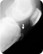
AN 18-year-old man complains of pain, swelling, and soreness of the right upper tibia. The pain is worse after playing basketball and climbing stairs. (Reprinted with permission from yochum TR, Rowe LJ. Essentials of skeletal radiology. 2nd ed. Baltimore, MD: Lippincott Williams & Wilkins; 1996:1291, Fig. 13.59B.)
CLINICAL POINTS
- The tibia tubercle(s) may appear prominent in long-standing cases.
- The disease usually begins in adolescence and resolves by adulthood.
- Symptoms may develop in association with growth spurts.
Clinical Presentation
Osgood–Schlatter disease is a common cause of knee pain in physically active adolescents. The disease is characterized by inflammation—pain, swelling, and tenderness—at the tibial tubercle. It is a nontraumatic condition. Patients commonly complain of pain and soreness at the tibial tubercle while climbing stairs. The tibial tubercle is the bony prominence below the patella; it is where the patella tendon is anchored. Clinical findings consist of pain, swelling, and tenderness of the tibia tubercle in an adolescent (or young adult) with normal stability tests. It is not uncommon for patients with chronic Osgood–Schlatter disease to develop a prominent tibia tubercle on one or both sides (Fig. 18.1). Approximately 25% of cases are bilateral.
Usually, Osgood–Schlatter disease resolves by early adulthood. The condition may be aggravated by sports that involve an extensive amount of running and jumping. Traditionally, the disease was thought to be more common in adolescent boys. However, it has been increasing in adolescent girls as well since athletic programs for girls have expanded.
The differential diagnosis of Osgood–Schlatter disease includes patellar tendonitis, prepatellar bursitis, infrapatellar bursitis, torn meniscus, torn cruciate ligament, torn patellar tendon, and pes anserine bursitis. Patellar tendonitis is characterized by pain and soreness of the patellar tendon with normal stability tests. Prepatellar bursitis is characterized by spongy swelling, tenderness, and soreness over the lower patella. Infrapatellar bursitis is seen as spongy swelling, tenderness, and soreness below the inferior pole of the patella. Pes anserine bursitis, is seen as focal tenderness of the pes anserine bursa. It is approximately 2 cm below the medial joint line.
Most patients with a torn medial meniscus have trouble squatting and are not able to duckwalk (walk in a squatting position). McMurray test can be used to confirm the presence of a meniscus tear; however, the sensitivity of the test is on the order of 50% to 60%. This test is performed with the patient supine by flexing the knee and then rotating the lower leg externally. Pain occurring along the medial joint line with external rotation that follows gradually straightening of the knee is positive for a medial meniscus tear. Pain that occurs along the lateral joint line with internal rotation followed by gradually straightening the knee is consistent with a lateral meniscus tear.
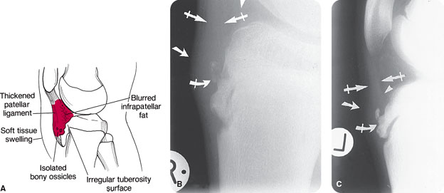
FIGURE 18.1 Characteristic features of Osgood–Schlatter disease. (A) Diagram. (B and C) Lateral knee. Observe the edema over the tibial tuberosity (arrows), blurred infrapatellar fat (arrowhead), thickened patellar ligament (crossed arrows), and fragmentation of the tuberosity (curved crossed arrows). (From Yochum TR, Rowe LJ. Yochum and Rowe’s Essentials of Skeletal Radiology. 3rd ed. Philadelphia, PA: Lippincott Williams & Wilkins; 2004.)
Related posts:


Stay updated, free articles. Join our Telegram channel

Full access? Get Clinical Tree


