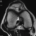
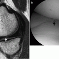
(1)
Department of Radiology, Saitama Medical University, Moroyama, Saitama, Japan
Abstract
Osteoarthritis (OA) is a joint disease represented by degeneration of cartilage, meniscus, subchondral bone, and other tissues due to ageing and mechanical load, abnormal proliferation of synovium, and bone and cartilage overgrowth.
10.1 Osteoarthritis
Osteoarthritis (OA) is a joint disease represented by degeneration of cartilage, meniscus, subchondral bone, and other tissues due to ageing and mechanical load, abnormal proliferation of synovium, and bone and cartilage overgrowth.
Of all joints of the body, the tibiofemoral joint of the knee is the most commonly affected site.
Common in elderly women.
Medial knee OA and varus deformity due to a loss of the medial joint space is more common than lateral knee OA.
Key points for MRI interpretation
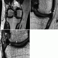
Joint space narrowing, osteophyte formation, sclerosis of subchondral bone, and cyst formation, all of which can also be detected by radiography. MRI-specific findings include articular cartilage thinning and loss, meniscal damage, and degeneration (Fig. 10.1).
Due to the high frequency of medial knee OA, deformity, and degeneration of the middle and posterior segments of the medial meniscus is frequently seen.
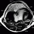
Joint effusion is commonly seen, reflecting the presence of cartilage and meniscal lesions (Fig. 10.2). Joint fluid in OA is clear, but MRI may show mucoid or mass-like signal changes within the joint space filled with effusion, representing secondary changes in a chronic stage. Proliferated synovium and denuded bone are prone to bleeding, and idiopathic hemarthrosis will recur in severe OA. In such cases, differential diagnoses include inflammatory arthritis and synovial disorders such as pigmented villonodular synovitis.
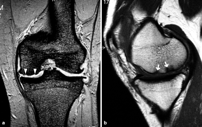
Fig. 10.1
Knee osteoarthritis. A woman in her 50s. (a) Coronal T2* WI and (b) PDWI. Medial joint space narrowing and the osteophyte formation are seen in the tibiofemoral joint. In the weight-bearing part, there is articular cartilage thinning (arrowheads), and subchondral sclerosis (arrows), degenerative tear, and loss of meniscus are seen
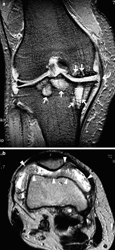
Fig. 10.2
OA and joint effusion. A woman in her 70s. (a) Coronal T2*WI and (b) axial T2WI. There are numerous subchondral cysts mainly in the medial tibiofemoral joint (arrows). MCL is extended in a bow shape due to the presence of medial osteophytes (curved arrow). Medial meniscus is severely degenerated and is almost completely macerated, but the medial joint space is not completely lost. A large amount of joint effusion is present together with proliferated synovium (arrowheads). Differential diagnosis may include synovial disorders such as pigmented villonodular synovitis in certain cases
Reference


Hayashi D, Guermazi A, Crema MD, Roemer FW. Imaging in osteoarthritis: what have we learned and where are we going? Minerva Med. 2011;102:15–32.
Intra-articular loose body
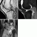
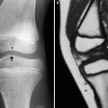
In chronic OA, ossified loose bodies may be found within the joint (Fig. 10.3)
It may appear as if it is continuous with an osteophyte (bony spur)
Loose bodies may freely move within the joint and may become stuck in a joint space, causing pain and limitation of range of joint movement.
Intra-articular loose bodies may also be associated with chronic arthritis, OCD, osteochondral fracture, and synovial osteochondromatosis.
Stay updated, free articles. Join our Telegram channel

Full access? Get Clinical Tree


