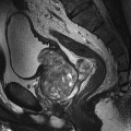
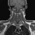
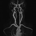
2 This section refers mainly to the Technical issues subheading discussed under the Image optimization heading considered for each examination in Part 2. Only a brief overview is provided here. For a more detailed explanation, please refer to Chapter 4 of MRI in Practice or an equivalent text. The main considerations of image quality are: Each factor is controlled by certain parameters, and each ‘trades off’ against the other (see later in Table 2.2). This section summarizes the parameters available and the trade-offs involved. Suggested parameters are outlined in Table 2.1, which can be found here and at the beginning of each anatomical region in Part 2. The parameters given should be universally acceptable on most systems. However, weighting parameters in particular are field strength dependent, and therefore, some modification may be required if you are operating at extremely low or high field strengths. Table 2.1 Summary of parameters The figures given are for 1.5 T and 3 T systems. Parameters are dependent on field strength and may need adjustment for very low or very high field systems. Table 2.2 Parameters and their trade-offs
Parameters and trade-offs
Introduction
1.5 T
3 T
SE
SE
Short TE
Min–30 ms
Short TE
Min–15 ms
Long TE
70 ms+
Long TE
70 ms+
Short TR
600–800 ms
Short TR
600–900 ms
Long TR
2000 ms+
Long TR
2000 ms+
FSE
FSE
Short TE
Min–20 ms
Short TE
Min–15 ms
Long TE
90 ms+
Long TE
90 ms+
Short TR
400–600 ms
Short TR
600–900 ms
Long TR
4000 ms+
Long TR
4000 ms+
Short TEL
2–6
Short TEL
2–6
Long ETL
16+
Long ETL
16+
IR T1
IR T1
Short TE
Min–20 ms
Short TE
Min–20 ms
Long TR
3000 ms+
Long TR
300 ms+
TI
200–600 ms
TI
Short or null time of tissue
Short ETL
2–6
Short ETL
2–6
STIR
STIR
Long TE
60 ms+
Long TE
60 ms+
Long TR
3000 ms+
Long TR
3000 ms+
Short TI
100–175 ms
Short TI
210 ms
Long ETL
16+
Long ETL
16+
FLAIR
FLAIR
Long TE
80 ms+
Long TE
80 ms+
Long TR
9000 ms+
Long TR
9000 ms + (TR at least 4 × TI)
Long TI
1700–2500 ms (depending on TR)
Long TI
1700–2500 ms (depending on TR)
Long ETL
16+
Long ETL
16+
Coherent GRE
Coherent GRE
Long TE
15 ms+
Long TE
15 ms+
Short TR
<50 ms
Short TR
<50 ms
Flip angle
20–50°
Flip angle
20–50°
Incoherent GRE
Incoherent GRE
Short TE
Minimum
Short TE
Minimum
Short TR
<50 ms
Short TR
<50 ms
Flip angle
20–50°
Flip angle
20–50°
Balanced GRE
Balanced GRE
TE
Minimum
TE
Minimum
TR
Minimum
TR
Minimum
Flip angle
>40°
Flip angle
>40°
SSFP
SSFP
TE
10–15 ms
TE
10–15 ms
TR
<50 ms
TR
<50 ms
Flip angle
20–40°
Flip angle
20–40°
Slice thickness 2D
Slice thickness 3D
Thin
2–4 mm
Thin
<1 mm
Medium
5–6 mm
Thick
>3 mm
Thick
8 mm
FOV
Matrix
Small
<18 cm
Coarse
256 × 128 / 256 × 192
Medium
18–30 cm
Medium
256 × 256 / 512 × 256
Large
>30 cm
Fine
512 × 512
Very fine
>1024 × 1024
NEX/NSA
Slice number 3D
Short
1
Small
<32
Medium
2–3
Medium
64
Multiple
>4
Large
>128
PC-MRA 2D and 3D
TOF-MRA 2D
TE
Minimum
TE
Minimum
TR
25–33 ms
TR
28–45 ms
Flip angle
30°
Flip angle
40–60°
VENC venous
20–40 cm/s
TOF-MRA 3D
VENC arterial
20–40 cm/s60 cm/s
TE
Minimum
TR
25–50 ms
Flip angle
20–30°
Parameter
Advantages
Disadvantages
TR increased (up to 2000 ms in SE)
Increased SNRIncreased number of slices per acquisition
Increased scan timeDecreased T1 weighting
TR decreased (below 2000 ms in SE)
Decreased scan timeIncreased T1 weighting
Decreased SNRDecreased number of slices per acquisition
TE increased
Increased T2 weighting
Decreased SNR ![]()
Stay updated, free articles. Join our Telegram channel

Full access? Get Clinical Tree

 Get Clinical Tree app for offline access


