George M. Bridgeforth and John Cherf
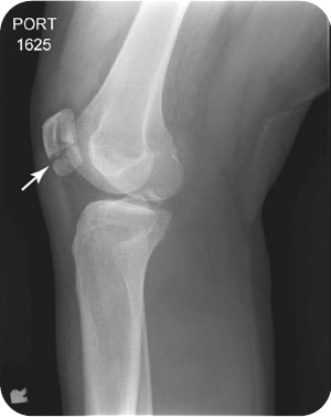
A 24-year-old woman presents with marked swelling, pain, and soreness of the right knee after a motor vehicle accident. She cannot bend or straighten her right knee.
CLINICAL POINTS
- Moderate to severe swelling, usually moderate to severe patella tenderness, is characteristic.
- Patients present with pain, limited range of motion, and antalgic gait (limp).
- Causes include direct impact and indirect blows.
Clinical Presentation
Patella fractures make up approximately 1% of all fractures and are usually seen in active people aged between 20 and 50 years. This injury is nearly twice as likely to occur in men compared with women. The mechanism of injury is usually a direct blow to the patella, resulting from a low-energy fall or a high-energy dashboard injury, or it may be indirect, resulting from running or jumping. Direct injuries cause comminuted fractures and indirect injuries, cause avulsion fractures, generally from the sudden contraction of the quadriceps.
Fractures are usually characterized by the acute onset of pain, swelling, and tenderness. There may be a large effusion, and patients are often unable to ambulate.
The physical examination of an acute injured knee may be hampered by apprehension and guarding. The initial inspection should check for signs of an effusion, acute inflammation, open wounds (potential portals for serious infections), or structural abnormalities. It should include an inspection of the entire leg for any evidence of a potential compartment syndrome (pallor or a swollen extremity). The presence of a large effusion indicates possible internal derangement from an intra-articular fracture or cruciate ligament tears. It should be noted that most knee fractures and contusions are not characterized by marked inflammation. On the other hand, septic joints have marked erythema, warmth, and tenderness. Untreated, septic joints may cause widespread joint destruction. Therefore, they should be subject to an immediate diagnostic aspiration; treatment is directed toward the underlying cause.
It is important to palpate the entire knee carefully for pain, swelling, and tenderness. Palpation should include not only the patella but also the joint spaces, the distal femoral condyles, the proximal tibia condyles (the epicondylar regions), the quadriceps and patella tendon, the pes anserine bursa, and the medial and collateral ligaments. Pain in the condylar regions may represent a condylar fracture, whereas pain along the medial joint line may be secondary to a meniscus tear. Focal tenderness of the pes anserine bursa about 2.5 cm below the medial joint line may be secondary to pes anserine bursitis. Although this remains a diagnosis of exclusion, it may be treated with analgesic medication and cold packs—a steroid injection may be reserved for refractory cases. The Ottawa rules concerning radiography (see “Radiographic Evaluation”) do not exclude underlying soft tissue injuries (i.e. tears); therefore, a thorough and systematic knee examination is essential.
Although the differential diagnosis for acute knee injuries is long, it is necessary to rule out a soft tissue contusion, an associated knee fracture, or internal derangement. A soft tissue contusion is characterized by pain, swelling, and tenderness. Although range of motion may be affected, stability of the knee is intact. Moreover, radiographs do not reveal any acute fractures. Follow-up studies such as a computed tomography (CT) or magnetic resonance imaging (MRI) should be reserved for patients who do not improve with conservative treatment (analgesics, cold packs, and physical therapy for severe cases). CT is slightly more sensitive for detecting an occult fracture, whereas an MRI is superior for detecting internal derangement (torn menisci, torn cruciate ligaments). However, MRI scans provide excellent sensitivity for detecting occult fractures.
Serious soft tissue injuries of the knee may occur following acute trauma, with or without an associated fracture. Twisting injuries may occur prior to impact, especially with falls and sports injuries. A torn medial meniscus (cartilage) is more common than a torn lateral meniscus. The medial meniscus is responsible for bearing a greater load (i.e., body weight), and it is firmly attached to the medial collateral ligament. Patients often complain of a burning pain along the inside of the knee—at the medial joint line. Associated complaints include trouble bending the knee, locking, clicking, and trouble using the stairs. Patients with torn cartilage have trouble performing and maintaining a full squat.
The following tests may be useful in examining the patient with a possible patella fracture:
- Straight leg raise: This test is used to detect disc injuries in the back (radicular pain past the knee from 30–60 degrees) or hip pathology (pain in the groin or hip at 30 degrees). There is an inability to extend the leg if the patellar tendon or quadriceps tendon is ruptured. If the patient has a torn meniscus, the knee may lock and the examiner cannot fully straighten the leg. But a straightened leg does not rule out a torn meniscus.
- McMurray test: This test is positive in patients with a torn medial meniscus. The knee is flexed and the lower extremity externally rotated with one hand while inward pressure is applied on the knee by the opposite hand. A positive test is characterized by pain or a painful clicking along the medial joint line.
- Apley compression test: This test is used to detect a torn meniscus only. It is performed with the patient lying face down. The knee is flexed to 90 degrees. Pain with compression and rotation along the joint line indicates a possible meniscal tear. Pain with distraction (as the lower leg is pulled upward) relieves the tension on the cartilage (menisci) and places a strain on the medial or collateral ligaments. Soreness with distraction coupled with palpable pain and tenderness along with good stability favors a medial or collateral ligament strain.
In addition to checking for meniscal injuries, it is important to check the structural integrity of the knee by checking for (ligament) instability. Patients with a completely torn medial collateral ligament exhibit valgus laxity. The unstable knee joint opens on the medial side as the examiner stabilizes the though with one hand as the lower extremity is gently pulled outward. Varus instability is caused by lateral collateral ligament injuries. The unstable knee joint opens on the lateral side as the lower extremity is gently brought inward by the examiner. Because the knee has an anatomic stabilization mechanism in full extension (called the “screw home” mechanism), varus and valgus testing should be performed when the knee is unlocked at 30 degrees. Some examiners have recommended repeating the testing when the knee is fully extended as well.
Patients with torn anterior cruciate ligaments (ACLs) often complain of a “pop” followed by knee pain and swelling. ACL tears may be seen with tibial spine or Segond fractures these. Associated fractures may or may not be present. ACL injuries should be suspected in patients with fractures of the tibial spine (median eminence) or with a Segond fracture. A Segond fracture is an avulsion fracture that occurs at the upper aspect of the lateral tibial plateau. It is best appreciated on the anteroposterior (AP) view. Moreover, Segond fractures have a very strong association with ACL or meniscus tears.
Several clinical tests are used to check for ACL tears. A Lachman test is more sensitive than an anterior draw test. With acute knee trauma, it may be difficult for patients to bend their knees, a Lachman test may be easier to perform as well. The patient flexes the knees to approximately 20–30 degrees some examiners perform the test with the knee slightly related externally. The examiner stabilizes the distal femur with one hand while placing the other hand on the proximal tibia. While keeping the knee stabilized, the tibia is pulled in an anterior direction (anterior translational force); the injured knee is compared with the opposite knee for evidence of joint laxity. A positive test is characterized by the anterior displacement of the tibia (>3 mm) that was not present on the opposite side. The anterior draw test is performed in a similar fashion, except that the knee is flexed to 90 degrees. Both hands are cupped firmly around the calf muscles (with both thumbs placed anteriorly over the knee) as the examiner tries to pull the tibia forward. Any detected laxity should be compared with the opposite knee. In addition, posterior cruciate tears may be detected by posterior translation (i.e., posterior laxity) of the tibia when pushing in the opposite (i.e., backward) directions.
PATIENT ASSESSMENT 
- Look for an associated effusion, which may or may not be present.
- Be sure to palpate the knee carefully.
- Determine whether any serious soft tissue injuries are present.
Related posts:


Stay updated, free articles. Join our Telegram channel

Full access? Get Clinical Tree


