(1)
Department of Radiology, Saitama Medical University, Moroyama, Saitama, Japan
Abstract
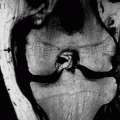
>Irregular appearance of the distal femur on radiograph.
9.1 Distal Femoral Cortical Irregularity
Irregular appearance of the distal femur on radiograph.
On anteroposterior radiograph, there is a round lucency within the femoral metaphysis together with sclerotic changes around it. On lateral radiograph, there is irregularity of the posterior femoral surface which mimics the periosteal reaction. In the past, malignant lesions were suspected and biopsy performed on these lesions.
In old days, it was also called “cortical desmoid.”
In fact, this is a bony irregularity due to traction by the medial head of gastrocnemius.
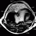
The attachment site of the adductor magnus is in the close proximity and may also be a cause for this lesion. The lesion will be located more medially in this case (Fig. 9.1).
Mostly bilaterally present in young persons, but may also be found in adults.
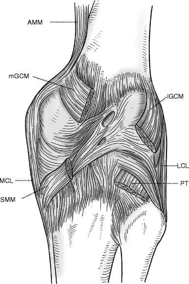
Fig. 9.1
The attachment sites of the medial head of gastrocnemius and adductor magnus. (Posterior view of the popliteal fossa.) The medial head of gastrocnemius (mGCM) and adductor magnus (AMM) attach to the medial side of the distal femur in the close proximity to each other. Distal femoral cortical irregularity occurs at these sites. lGCM lateral head of the gastrocnemius, PT popliteus tendon, SMM semimembranosus, MCL medial collateral ligament, LCL lateral collateral ligament
Key points for MRI interpretation
An area of hypointensity representing sclerotic changes of the bone and varied signal changes within it (Fig. 9.2).
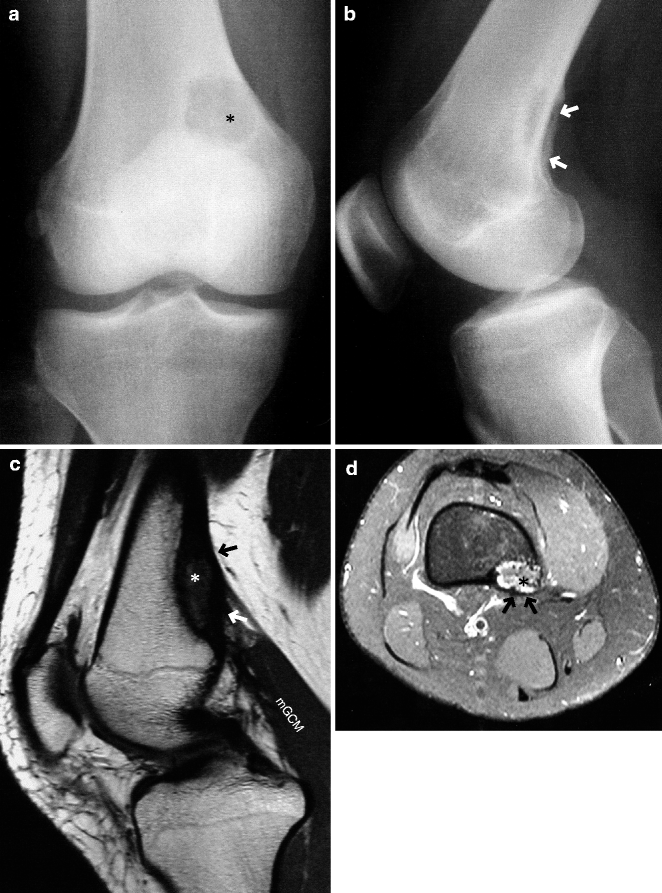
Fig. 9.2
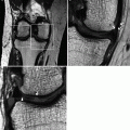
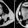
Distal femoral cortical irregularity. A woman in her late teens. (a) Anteroposterior and (b) lateral radiographs. (c) PDWI and (d) axial FS T2WI. There is a round lucency in the distal femoral metaphysis (*, a) and the surrounding sclerotic changes on anteroposterior radiograph. Lateral radiograph shows protruding irregularity of the posterior femur (arrows). On MRI, near the attachment site of the medial head of gastrocnemius (mGCM), there is a band of hypointensity corresponding to the sclerotic changes and varied signal intensities within it (*). Similar changes were also in the contralateral femur
References
Resnick D, Greenway G. Distal femoral cortical defects, irregularities, and excavations. Radiology. 1982;143:345–54.
Bufkin WJ. The avulsive cortical irregularity. Am J Roentgenol Radium Ther Nucl Med. 1971;112:487–92.
9.2 Femoral Condylar Irregularity
Commonly seen in children who are 2–6 years old (in more than 40% of cases). Also common in teenagers.
May be an incidental finding on radiograph, and the patient may then be sent to have an MRI (Fig. 9.3).
More common in the lateral femoral condyle (40% of cases involve the lateral condyle only) but can also occur in the medial condyle (Fig. 9.4).
Commonly seen in the posterior side of the femoral condyle (Fig. 9.5). (cf. Osteochondritis dissecans (OCD) commonly occurs in the lateral side of the medial femoral condyle. See Chap. 8 for differential diagnosis of OCD and spontaneous osteonecrosis.) Therefore, the lesion may not be visible on anteroposterior radiograph. Tunnel view radiograph may be useful.
Articular cartilage is normal.
Patients are asymptomatic and the irregularity disappears after a few years (Fig. 9.6).
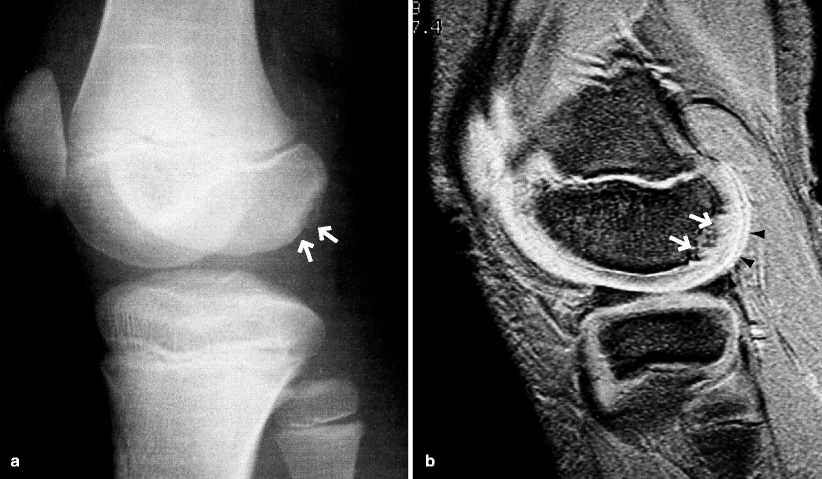
Fig. 9.3
Femoral condyle irregularity. An 11-year-old boy. (a) Oblique view radiograph and (b) T2*WI. There is a minute irregularity of the posterior aspect of the lateral femoral condyle (arrows) on radiograph. On MRI, there is irregularity of cortical bone (arrows), but the articular cartilage is absolutely normal (arrowheads). Similar changes were also seen in the contralateral knee
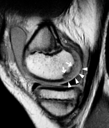
Fig. 9.4
Femoral condyle irregularity of the medial femoral condyle. A 10-year-old boy. PDWI. There is irregularity of cortical bone in the posterior aspect of the medial femora condyle (arrows), but the articular cartilage is absolutely normal (arrowheads)
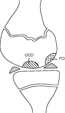
Fig. 9.5
Common sites of occurrence of the femoral condyle irregularity ( FCI ) and osteochondritis dissecans ( OCD ) (Schematic illustration of a sagittal knee image in a young person)
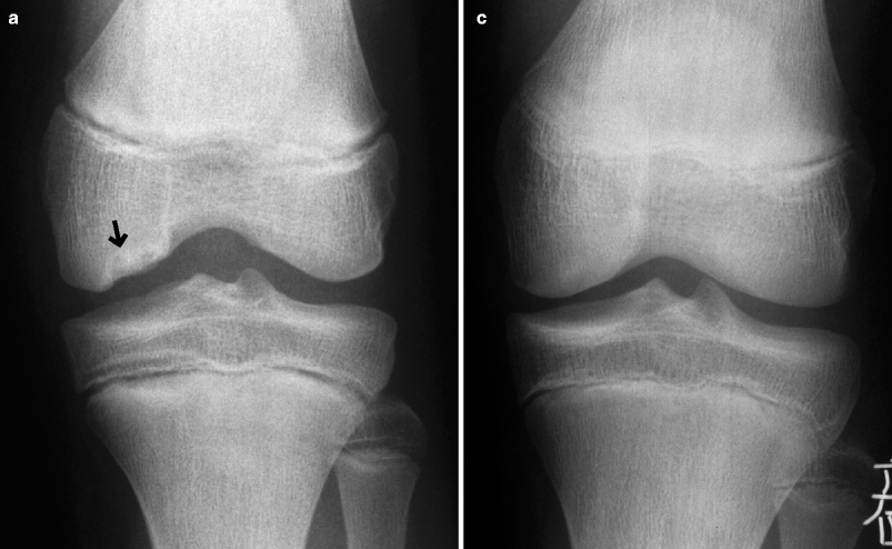
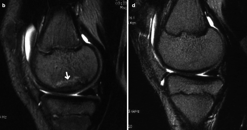
Fig. 9.6
Femoral condyle irregularity that disappeared after 2 years. A 12-year-old boy (at the time of initial diagnosis). (a) Tunnel-view radiograph and (b) T2WI at the time of presentation. (c) Tunnel-view radiograph and (d) T2WI 2 years later. There is a femoral condyle irregularity in the weight-bearing portion of the medial femoral condyle (arrows). However, the lesion disappeared 2 years later (Image courtesy of Dr. Shigeru Ehara, Department of Radiology, Iwate Medical University, Japan)
Key points for MRI interpretation
The site of the lesion is the key for correct diagnosis. Imaging appearance may be similar to OCD, but the site of lesion is different. Diagnosis of femoral condylar irregularity can be confirmed by the presence of normal cartilage on MRI.
Reference
Caffey J, Mandell SH, Royer C, et al. Ossification of the distal femoral epiphysis. J Bone Joint Surg. 1958;40-A:647–54.
9.2.1 Disorders Characteristics to Knees of Children and Adolescents
There are painful knee disorders that characteristically occur in children and adolescents, commonly due to overuse of the bone, cartilage, and ligaments which are subjected to excessive mechanical stress following strenuous sports activities. Such disorders include Osgood-Schlatter disease and Sinding-Larsen-Johansson disease, both of which are thought to be osteochondrosis.
9.3 Painful Patella Partita
Patella partita is a malformation of the patella which comprises more than one part.
It is thought to be caused by incomplete ossification process of two or more ossification centers, or by excessive stress from sports, but it is considered to be a normal variant. Bilateral knees are affected in approximately half the cases. Most cases are thought to naturally heal before the adolescence.
More common in boys.
A classification system developed by Saupe is commonly used (Fig. 9.7). Saupe type III is the most common (Fig. 9.8) because of poor vascular supply in the superolateral pole of the patella, to which the vastus lateralis attaches.
Patella bipartita has two parts (Fig. 9.8
Stay updated, free articles. Join our Telegram channel

Full access? Get Clinical Tree


