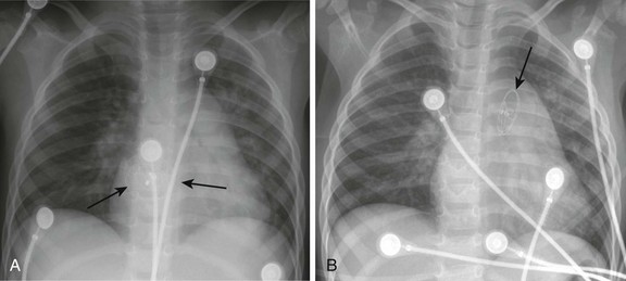
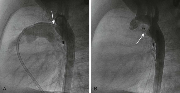
Pediatric Cardiac Catheterization and Electrophysiology

Chapter 69


Pediatric Cardiac Catheterization and Electrophysiology


Pediatric Cardiac Catheterization


Procedural Techniques


Catheterizations are performed with use of the standard Seldinger technique in the femoral artery and/or vein. Alternative venous access includes the internal jugular, subclavian, or transhepatic approaches. Arterial access also may be obtained via the arteries of the upper extremities or the carotid artery, but these approaches typically are restricted to larger patients or emergent conditions. Anticoagulation is managed with heparin, 80 to 100 U/kg or 5000 U in patients who weigh more than 50 kg, for an activated clotting time goal of 200 to 250 seconds, depending on the procedure to be performed. Antibiotics are administered to patients who receive an implanted device. Baseline hemodynamics (including pressures and oxygen saturations) and angiography are obtained in room air, whenever possible. Subsequently, relative blood flows and vascular resistances are calculated. The ratio of pulmonary blood flow (Qp) to systemic blood flow (Qs) is calculated according to the following formula: Qp/Qs = [(Ao2 − Mvo2)/(Pvo2 − Pao2)], where Ao2 is the aortic oxygen saturation, Mvo2 is the mixed venous oxygen saturation, Pao2 is the pulmonary arterial saturation, and Pvo2 is the pulmonary venous saturation. These data then determine whether additional information or interventions are necessary.


Semilunar Valve Stenosis and Balloon Valvuloplasty


Since transcatheter balloon pulmonary valvuloplasty for valvar pulmonary stenosis in infants was first reported in the early 1980s, it has become the first line of therapy. The recommended balloon/annulus diameter ratio is 120%.2 The reported success rate is greater than 90%, with major adverse events occurring in fewer than 1% of the procedures.3 Hemodynamic measurements include the right ventricular pressure compared with systemic arterial pressure and the peak-to-peak systolic pressure gradient across the pulmonary valve. The indication for balloon pulmonary valvuloplasty is the presence of at least moderate pulmonary valve stenosis. We use as a guideline the “rule of 50,” which is defined as a peak right ventricular systolic pressure of more than 50 mm Hg, a peak right ventricular systolic pressure more than 50% of the systemic systolic pressure, or a peak-to-peak systolic gradient across the pulmonary valve of more than 50 mm Hg.


Valvar aortic stenosis can be classified into two groups: disease that is severe enough that it presents at birth or within 1 year of age (10% to 15%), and disease that is not diagnosed until after age 2 years and will progress much more slowly, if at all.4,5 Mortality and the need for intervention are significantly skewed toward the infantile group. As with pulmonary stenosis, noninvasive imaging techniques have advanced to the point that nearly all anatomic and functional information about the valve may be obtained without catheterization. Catheterization is performed for valves that clearly merit intervention or when symptoms and imaging findings are incomplete or confounding.


Aortic valve stenosis is classified into the following categories: trivial, mild, moderate, severe, and critical. Critical aortic stenosis is not defined by a specific pressure gradient or valve orifice size but on the basis of physiologic manifestations. If the stenosis is such that the patient is unable to produce and maintain an adequate cardiac output, the stenosis is critical. Patients in this group may have a low valve gradient, as measured by echocardiography, because of decreased cardiac function and low cardiac output. Although some controversy still exists with regard to the most beneficial treatment method for this population (i.e., surgical valvotomy vs. percutaneous balloon valvuloplasty), most centers have adopted balloon valvuloplasty as the initial treatment of choice. Patients in this category do not tolerate the stress of any procedure well, and catheterization has immediate results comparable with those of surgery (i.e., a reduction in gradient and resultant valve regurgitation) with a shorter course in the intensive care unit after the procedure and a shorter overall hospital stay.


Balloon valvuloplasty has been associated with an increased rate of reintervention compared with surgical valvotomy as a result of recurrent stenosis or worsening regurgitation. Given that residual aortic valve disease, especially regurgitation, may progress over time, the recommendation for the valvuloplasty technique is more conservative, with a smaller maximal balloon diameter (80% to 100% of the annulus) than that recommended for the pulmonary valve (100% to 120%). The valve may be approached retrograde from the aorta, using a soft-tipped J-wire to cross the narrowed valve orifice and obtaining arterial access in the femoral artery (more commonly) or the carotid artery. The valve also may be approached prograde by crossing an existing atrial communication or by performing a transseptal puncture to access the left side of the heart. Once in the left ventricle, angiography is performed to measure the annulus of the valve and obtain landmarks for valvuloplasty. The diameter of the balloon should not exceed 80% to 90% of the valve annulus. The smaller balloon diameter, compared with a similar sized pulmonary valve annulus, is recommended to decrease the amount of valve tearing and resultant acute regurgitation.


Many centers have adopted rapid right ventricular pacing at the time of balloon inflation. This rapid pacing transiently reduces cardiac output and the shearing force transmitted to the balloon as it is inflated across the valve annulus. The goal is to reduce the motion on the fragile valve leaflets and prevent excessive damage and regurgitation. Repeat angiography and echocardiography after inflation are essential to evaluate the success of the valvuloplasty and monitor for regurgitation or other complications.


The differentiation between noncritical stenosis categories is made by noninvasive echocardiographic measurements of valve area and Doppler gradient. A normal valve area is 2 cm2/m2. Mild obstruction is consistent with valve areas less than 2 cm2/m2 but greater than 0.7 cm2/m2, and severe obstruction is consistent with valve areas less than 0.5 cm2/m2. Mean echocardiographic Doppler gradients are good predictors of the peak-to-peak pressure gradient measured at catheterization. Gradients less than 25 mm Hg are considered trivial, those 25 to 50 mm Hg are mild, those 50 to 75 mm Hg are moderate, and those >75 mm Hg are severe. These measurements are made with the understanding that the cardiac function and cardiac output are normal.


Catheterization is not recommended for trivial or mild stenosis. Moderate and severe stenoses are approached with primary balloon valvuloplasty using the techniques previously described.


Balloon or Stent Angioplasty


Since the late 1980s, when balloon angioplasty was performed with low-pressure balloons (<5 atmospheres), balloons capable of dilations at higher pressure have been introduced. Since the introduction of high-pressure balloons, standard balloon angioplasty has resulted in a successful result of more than 70% for pulmonary artery angioplasty. As for coarctation or recoarctation, balloon angioplasty also has been used with some success. Patients who have severely hypoplastic pulmonary arteries with multiple stenoses that are refractory to high-pressure angioplasty may not be treatable using currently available medical, surgical, or catheter-based tools. In such cases, a “waist” persists during angioplasty at the maximum recommended inflation pressure, or even at pressures exceeding the recommended maximum, because of inadequate stretching or tearing of the vessel wall. For this patient population, cutting balloon angioplasty has become a therapeutic option with use of balloons up to 8 mm in diameter. Cutting balloon angioplasty often is followed by standard angioplasty for the best final result. Balloon-expandable stents have improved results in proximal vessels, eliminating the need for surgery in most patients, but they are of limited value in distal pulmonary vessels. Furthermore, stents are contraindicated in noncompliant vessels that cannot be expanded using high-pressure balloons. Stent angioplasty also has been utilized for coarctation of the aorta in postoperative and native lesions.


Pulmonary artery stenosis is a form of congenital heart disease that can occur in isolation or as part of more complex malformations such as tetralogy of Fallot. Although obstructions confined to the main or proximal branches can be repaired surgically, many patients can receive adequate palliation with transcatheter balloon angioplasty techniques. In addition, more distal obstructions that cannot be repaired operatively require angioplasty with balloons delivered to the distal stenotic segments as previously described. The inflation of a balloon whose maximum diameter is two to three times the diameter of the lesion will tear the vessel wall within the stenotic segment, resulting in an increase in lumen diameter. Although balloon catheters have not specifically been approved by the Food and Drug Administration as a treatment for pulmonary artery stenoses, transcatheter techniques have become the mainstay of treatment for distal vessel obstruction.


Often, aortic obstruction after an end-to-end surgical repair at the isthmus is a result of aortic narrowing within the transverse arch. These obstructive lesions are further defined as either the proximal transverse arch between the innominate artery and left carotid artery or the distal transverse arch, defined as the region between the left carotid artery and the left subclavian artery. Although few data exist regarding stent angioplasty within the distal transverse aortic arch, general experience has been that this procedure is safe and effective. An arterial monitoring catheter is placed in the right upper extremity and a 4 Fr sheath is used to advance a 4 Fr pigtail to the aorta from the right radial artery. This catheter is used to monitor pressures during stent angioplasty of the distal transverse arch and to perform cine angiograms to determine appropriate stent placement distal to the takeoff of the left carotid artery.


Transcatheter stent angioplasty for postoperative recoarctation of the aorta at the isthmus has been demonstrated to be safe and effective.5 The technique usually involves a femoral arterial approach. An exchange length wire is placed in the ascending aorta or right subclavian artery. An angioplasty balloon is used with a maximum diameter that is equal to or less than the diameter of the normal aortic segments adjacent to the stenotic region and/or the diameter of the descending thoracic aorta at the diaphragm. The stent is mounted on the angioplasty balloon and passed through a sheath at least 1 to 2 Fr larger than that required by the balloon. The stent length is dependent on the lesion length but usually is at least 36 mm in adults. The stent is fully dilated in most cases, but at times it is deemed safer to serially dilate the lesion over two procedures.


Septal and Vascular Occlusion Devices


Although diagnosing the presence of an atrial septal defect (ASD) rarely requires cardiac catheterization, today many patients are undergoing cardiac catheterization for therapeutic device closure.6 These patients require assessment of associated anomalies such as abnormalities of pulmonary venous connections. A step-up in oxygen saturations in the right atrium and pulmonary arteries is characteristic for an ASD, and the degree of left-to-right shunting or the pulmonary to systemic blood flow ratio (Qp : Qs) can be determined. The ideal age or timing for elective ASD closure is 2 to 5 years of age or within 6 to 12 months of diagnosis. Rarely, a child with an ASD presents with severe congestive heart failure and requires intervention in the first year of life.


Percutaneous ASD closure has been established as a safe and effective alternative to operative repair. The technique involves transcatheter delivery of a device in its retracted state via the femoral vein under fluoroscopic and echocardiographic guidance. Echocardiographic guidance can be transthoracic in young children and transesophageal, intracardiac, or even three-dimensional (3D) in older children. The most commonly available devices today consist of a double disc design made of nickel and titanium (Nitinol) with deployment of the first disc on the left atrial aspect of the septum followed by deployment of the second opposing disc on the right atrial wall. The expanded discs are tightly approximated, thus closing the defect. The device becomes endothelialized during the next 3 to 12 months while the patient is treated with antiplatelet medications. Contraindications include some very large defects, the absence of adequate septal tissue margins, close proximity to vital cardiac structures, and very small children. ASD devices require an adequate tissue margin (minimum 7 to 8 mm) for deployment. The ASD must be small enough that the device can be deployed and held in the atrial septum (an adequate margin must be present on all sides other than the anterior superior rim by the aorta, where some splay of the device around the aorta will compensate for deficient rim) without impingement on adjacent structures or significant pressure on the walls of the atrium. Venous inflow to both the right and left atria must not be impeded, and the tricuspid and mitral valves should not come into contact with the device.


The world experience of transcatheter ASD closure using the Food and Drug Administration–approved Amplatzer Septal Occluder (AGA Medical Corporation, Golden Valley, MN) has been reported with a greater than 97% immediate success rate.7 The occlusion rate reached 100% by 3 years. An overall 2.8% adverse event rate was found, and no procedural deaths occurred. Several studies regarding comparisons of device occlusion versus surgical closure have been performed prospectively. These reports include the Amplatzer and Helex (W.L. Gore & Associates, Flagstaff, AZ) devices for ASD closure. Findings include shorter hospital stays, less discomfort, and shorter durations for convalescence in patients undergoing successful closure with use of a device. Hospital costs are similar. Regression of right ventricular dilatation was similar for both groups of patients; however, it was dependent on the patient’s age at the time of closure, with greater regression following earlier intervention.


Procedural adverse events are uncommon but include embolization into the right or left atrium, pulmonary artery, left ventricle, and aorta; stroke as a result of a clot or air embolization; and bleeding complications.8 Both acute and late embolization have been reported, and thus it is critical for the radiologist to be familiar with the location of the interatrial septum on radiography to ascertain correct device position in the anteroposterior and lateral chest radiograph. Figure 69-1, A, demonstrates a septal occluder device in the appropriate position within the interatrial septum, and Figure 69-1, B, demonstrates the device positioned incorrectly in the left pulmonary artery.


The first transcatheter interventional procedure was closure of a persistently patent ductus arteriosus (PDA), which was performed by Portsmann in 1967. Since that time a number of different PDA closure devices have been studied, some of which are no longer available. The goal of the procedure has always been to achieve 100% closure of the PDA without obstruction of either adjoining blood vessel (i.e., the aorta and the left pulmonary artery) with minimal risk of complications. The difficulty is that the PDA exhibits extreme variability in size and shape. Krichenko et al.9 classified the PDA into five anatomic types based on the lateral aortic angiogram. The most common (“type A”) ductus is conical with a narrowed pulmonary arterial end and large aortic ampulla. Other types include those with a narrowed aortic end, narrowing at both ends, and a tubular configuration. Early device closure procedures were complicated by a lack of choices for vessels of different sizes and shapes. The early devices had unsatisfactory rates of complications and residual leaks and were abandoned. In 1992, the first report was published using the Gianturco embolization coil (Cook Medical, Bloomington, IN) for closure of small PDAs. The Gianturco coil, which is available in multiple lengths and diameters, has now been in use for more than 20 years for blood vessel occlusion, including unwanted collateral vessels, fistulae, and arteriovenous malformations. The successful use of the coil in PDA closure, combined with sharing of ingenious techniques to deploy multiple coils and secure the coils before deployment by individual operators, provided the needed variety of approaches for successful PDA closure. At present, tens of thousands of patients around the world have had PDAs closed with embolization coils.


As expected, the recommended technique for coil embolization is variable, depending on the size and shape of the PDA. The vessel can be approached from the venous or arterial side, and the coils may be deployed “free hand” or secured with a bioptome or modified catheter. Smaller coils, such as the Flipper coil (Cook Medical), also are available; these coils are attached to a delivery system and are released once they are verified to be in the proper position.


A more recent addition to the interventional cardiologist’s armamentarium for PDA closure is the Amplatzer Duct Occluder.10 The PDA occluder is a self-expanding wire mesh device that is attached to a delivery cable and deployed through a long sheath from the venous system. Figure 69-2, A, demonstrates a lateral projection angiogram of the descending thoracic aorta and a left-to-right shunting PDA. Figure 69-2, B, demonstrates device occlusion of the PDA. The device has a retention skirt to occupy the aortic ampulla and tapers slightly to the pulmonary artery end. The device is filled with a polyester mesh that stimulates thrombus formation within the lumen of the PDA. The shape of the device and self-expanding properties exert radial force on the walls of the PDA, holding the device in place until endothelialization occurs. The device has excellent closure rates approaching 100% at 1 month after the procedure. The main limitation is the size and bulky nature of the device. The PDA must have an aortic ampulla adequate to accommodate the retention skirt without creating aortic obstruction. Furthermore, the device can create left pulmonary artery stenosis by compressing adjoining structures once it is released.


Accessory blood vessels are a common area of concern for the interventional radiologist and interventional cardiologist. These accessory blood vessels may include aortopulmonary or venovenous collaterals, arteriovenous fistulae and malformations, surgically placed shunts, and transhepatic access tracts. Techniques for closure are similar to those described for the PDA, predominately involving Gianturco and similar coils, the Amplatzer Duct Occluder, or the Amplatzer Vascular Plug. The vascular plug is similar to the duct occluder in that it is a self-expanding wire mesh design. It differs in that it is cylindrical in shape with no retention skirt, no tapering through its length, and no polyester mesh interior fabric. The device has excellent occlusion results but has been unsuccessful in short arterial vessels, such as aortopulmonary collaterals and the PDA. It is thought that the lack of occlusive material through the center of the device does not provide enough restriction to arterial blood flow to stimulate thrombosis and occlude the vessel.


Dec 20, 2015 | Posted by in PEDIATRIC IMAGING | Comments Off on Pediatric Cardiac Catheterization and Electrophysiology

Full access? Get Clinical Tree

Get Clinical Tree app for offline access