Patricia E. Burrows
BACKGROUND
An arteriovenous malformation (AVM) is characterized by the presence of direct arteriovenous connections leading to arteriovenous shunting. Several genetic mutations affecting the transforming growth factor-beta superfamily and the Ras signaling pathway are known to cause familial AVM, but most AVMs are sporadic. In the female pelvis, two sites account for the most AVMs: the pelvic wall and the uterus. These are often referred to as congenital and acquired AVMs, respectively, but neither is seen in early childhood.
Pelvic Wall Arteriovenous Malformation
Pelvic wall AVMs primarily involve the bone and soft tissue of the pelvic wall and drain into branches of the internal iliac veins. As they enlarge, arteries and veins from the pelvic viscera can be recruited. An AVM of the pelvic wall is typically supplied by numerous branches of the pudendal, obturator, and gluteal arteries, as well as collaterals from adjacent visceral branches, and can achieve massive size and flow. Although some asymptomatic patients are diagnosed by cross-sectional imaging of the pelvis for other indications, the majority present with pain, bleeding, dyspnea due to high-output heart failure, or combinations of these symptoms.1 Footdrop due to nerve compression and lower extremity edema due to associated proximal iliac vein compression have also been described.2 Ligation or proximal embolization of the internal iliac arteries is frequently performed but is always ineffective because of the rapid reperfusion of the AVM through collaterals.3
Uterine Arteriovenous Malformation
Uterine AVMs typically result from uterine trauma, such as pregnancy or curettage, or in association with trophoblastic malignancy and typically present with vaginal bleeding, sometimes years after the trauma. Uterine AVMs arise in the muscle of the uterus and typically are supplied by uterine and ovarian arteries and drain into gonadal and internal iliac veins. Extensive lesions can involve vessels from the adjacent bladder and vagina. Color Doppler ultrasonography is diagnostic. Retained products of conception should be ruled out.
TECHNIQUES
One suggested technique for embolization of arteriolovenous fistula or AVM with single venous outflow is as follows:
1. Perform a pelvic aortogram to delineate arterial and venous anatomy.
2. Perform a selective internal iliac angiogram to better define the vascular anatomy, as needed.
3. Identify the point at which all of the feeding arteries connect to the same segment of draining vein.
4. Access the immediate draining vein (transvenous or percutaneous). Percutaneous access is achieved with ultrasound guidance or fluoroscopic guidance with or without road map.
5. Deliver a detachable framing coil or other large occlusive device into the immediate draining vein and continue packing the varix with coils. When the coil mass is stable and has slowed the blood flow, ethanol or N-butyl cyanoacrylate (NBCA) can be injected into the coil mass or additional coils can be placed to reach occlusion. If the draining vein is massive, outflow control can be assisted by transjugular positioning of an occlusion balloon or partly deployed inferior vena cava (IVC) filter. These can be removed after the coil mass has been created and deemed to be stable.
6. Repeat internal iliac arterial or aortic contrast injections to monitor the occlusion of the venous outflow.
7. Small residual shunts can be occluded by transarterial microcatheter embolization or percutaneous direct puncture technique.
Techniques for embolization of uterine AVM:
1. Perform abdominal aortogram to identify arterial supply and angioarchitecture.
2. Perform selective, bilateral uterine artery catheterization.
3. For an AVM with small arteriovenous shunts, bilateral uterine artery embolization with large particles and/or Gelfoam (Pfizer, New York, New York) is carried out.
4. For an AVM with large arteriovenous shunts, superselective or percutaneous embolization using permanent occlusive agents such as NBCA, Onyx (Covidien, Irvine, California), or coils is used.
5. Ultrasound-guided percutaneous cannulation of nidus, or immediate draining vein, followed by intranidal embolization with ethanol, glue, or Onyx is also effective in achieving obliteration of the shunt.
6. Perform postembolization angiography.
7. Clinical follow-up supplemented with color Doppler ultrasonography.
OUTCOMES
Pelvic Wall Arteriovenous Malformation
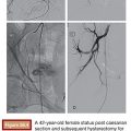
When treating pelvic wall AVMs, arterial embolization using acrylic embolization material, coils, or particles can result in symptomatic improvement but is rarely curative because of the large number of feeding arteries involved.4–6 Jacobowitz and colleagues5 reported a retrospective review of 35 patients undergoing arterial embolization, using acrylic polymer (glue), of pelvic vascular malformations; most were AVMs. Presenting problems included pain (59%), a visible or palpable lesion (62%), pulsation or thrill (44%), hemorrhage (27%), congestive heart failure (18%), and symptoms due to mass effect (35%). One-third of patients had undergone previous, unsuccessful attempted surgical treatment of the lesion. Patients underwent a mean of 2.4 embolization procedures over an average period of 23.3 months. Five patients went on to have adjunctive surgical procedures. Eighty-three percent of patients were asymptomatic or significantly improved at mean follow-up of 84 months, although none were cured, and symptomatic recurrences occurred frequently. Ethylene vinyl alcohol copolymer (Onyx) has been effective in a few cases because of the characteristic of this agent to progressively penetrate anastomotic feeding channels as well as the draining vein.7
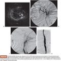
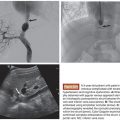
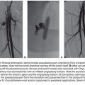
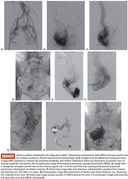
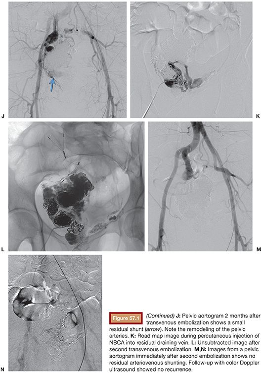
Because most pelvic AVMs have single outflow venous anatomy, they are best treated by outflow venous occlusion.1,3,4 In this technique, the outflow vein is packed with embolic material, usually coils, but also potentially ethanol or a polymerizing agent (NBCA, Onyx) (Fig. 57.1). This obviates, or markedly reduces, the need for and risks associated with superselective embolization of the pelvic arteries. The procedure involves careful review of arteriographic images to identify the common immediate draining vein. The immediate draining vein can be catheterized from the contralateral femoral vein, the internal jugular vein, or percutaneously using a micropuncture system or catheter introduced directly through the pelvic wall. My preference is to use the direct percutaneous approach because it is the quickest and most accurate method of catheterizing the immediate draining vein. Retrograde catheterization from the contralateral femoral vein is the most difficult because the draining pelvic veins are usually tortuous. Complications of venous embolization include pulmonary embolism and pelvic bleeding from residual arterialized, partly occluded veins. Migration of coils or thrombus can be controlled by proximal venous balloon occlusion, although this can be difficult because of the extremely large size of the draining vein. Placement of an IVC filter or other tethering device in the internal iliac vein can be effective in preventing dislodgment of coils.4 Fortunately, there are now many detachable coils available in large diameters. These can be used to create a frame or basket to which pushable, large-diameter coils can be added. My approach to the massive draining vein involves initial placement of coils into the varix, followed by injection of NBCA into the coil mass. Additional arterial or percutaneous embolization may be needed to complete the occlusion. In the largest published series of venous outflow occlusion for pelvic AVM, 12 patients underwent coil embolization of the dilated draining vein followed by transarterial ethanol embolization. Eighty-three percent of patients were cured with one major complication (focal bladder necrosis).1
Stay updated, free articles. Join our Telegram channel

Full access? Get Clinical Tree


