Pelvis, Including Lower Urinary Tract Trauma


Pelvis, Including Lower Urinary Tract Trauma


John H. Harris Jr.


GENERAL CONSIDERATIONS

In this chapter, the pelvis is considered to include the bony pelvis and its extraperitoneal soft tissues. Injuries involving the pelvis and its contents result in some of the most challenging and serious diagnostic problems to confront the radiologist, emergency physician, and traumatologist. Radiographically, this is particularly true for two reasons. First, the pelvis is the single area of the body that defies the radiologic dictum of obtaining radiographs in both frontal and lateral projections. Second, the soft tissue injuries frequently associated with pelvic skeletal injury are radiographically much less striking than the skeletal injury but are of far greater clinical significance.

The principal cause of death associated with pelvic trauma is hemorrhage, estimated to be as high as 60% in patients with major pelvic trauma.1, 2 Interventional angiography has resulted in a significant reduction in this mortality rate.3, 4

Rupture of the bladder or urethral injury occurs in approximately 20% of patients with significant pelvic ring disruption (PRD). However, bladder and urethral injuries should be suspected in all patients with major pelvic trauma.5, 6, 7

The proper evaluation of urethral injuries requires a retrograde urethrogram (RUG) as the initial diagnostic procedure in all male patients with a PRD who are unable to void spontaneously.8, 9, 10, 11, 12, 13, 14, 15 If the RUG is negative, a cystogram should be performed next. There is no justification for the insertion of a Foley catheter before performing an RUG in male patients with PRD. All male patients suspected of having a urethral injury, either on a clinical basis or because of radiographically demonstrated PRD, must have an RUG prior to insertion of a Foley catheter.


RADIOGRAPHIC ANATOMY

It is important to be familiar with changes in the radiographic appearance of the pelvis related to normal growth and development. This is so that (1) normal anatomy may not be misinterpreted as signs of trauma and (2) the basis of avulsive injuries of the adolescent pelvis may be understood and recognized.

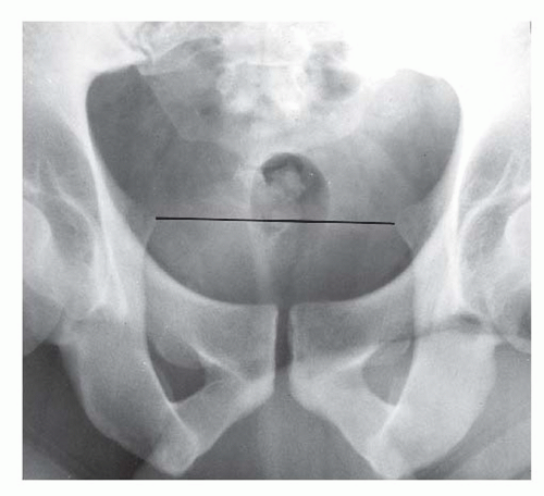
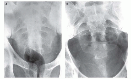
The radiographic appearance of the pelvis of a normal child is seen in 
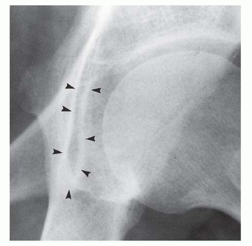
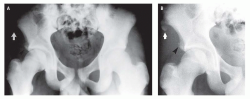
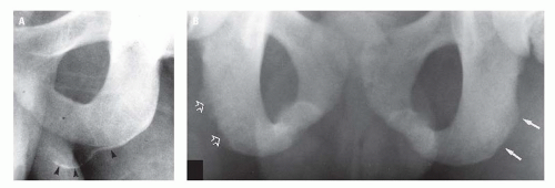
Figure 17.6. The triradiate cartilage (TRC), the site of union of the pubis, ischium, and ilium, usually fuses concomitant with puberty. The ischiopubic synchondrosis may fuse between the ages of 5 and 12 years.
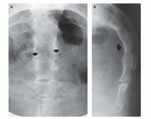
19 This synchondrosis varies greatly in radiographic appearance during the fusion process, not only from child to child (Fig. 17.7) but also from side to side in the same child (Fig. 17.8). The appearance of this synchondrosis rarely suggests an acute fracture but very commonly simulates a healing one (Fig. 17.8). By age 14 years, the TRC is nearly fused and the ischiopubic synchondroses are fused (Fig. 17.9).


Figure 17.6. AP radiograph of the pelvis of a normal 8-year-old child. Normally in children, the SI joints (open arrow) appear abnormally wide because of incomplete ossification of the contiguous surfaces of the SI joints. The triradiate cartilages (white arrow) are physiologically open until ages 13 to 16 years. Also, by this age, the ischiopubic synchondroses (black arrow) are fused.


Figure 17.7. A and B: Examples of the variability of the radiographic appearance of the ischiopubic synchondrosis in children of different ages. In the younger child (A), the right synchondrosis (arrow) could be misinterpreted as either an acute incomplete or a healing fracture.

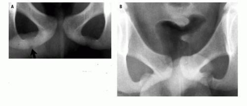
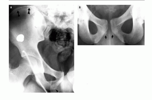
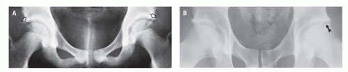
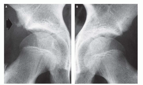
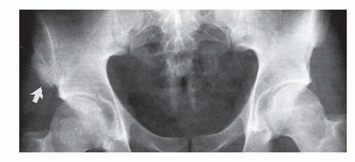
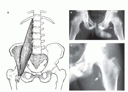
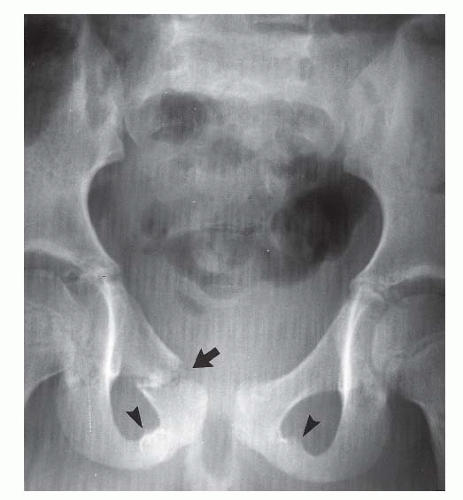
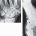
The pelvis of adolescents and young adults contains several apophyses that may be mistaken for acute fractures or that may be the site of acute avulsive injuries. These include apophyses of the iliac crest, the anterosuperior and inferior iliac spines, the ischial tuberosity (Fig. 17.10), and the inferior margin of the pubic bodies (Fig. 17.10B), all of which ossify with puberty and fuse between the ages 20 and 25 years.20 The anterosuperior iliac spine apophysis is the site of origin of the sartorius muscle; the anteroinferior iliac spine apophysis, the rectus femur muscle; and the ischial apophysis, the hamstring muscles.

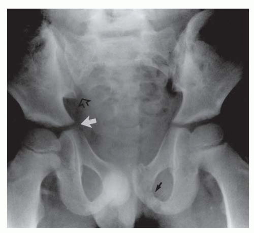
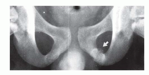
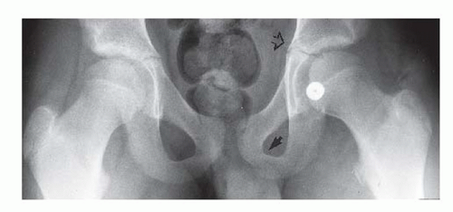
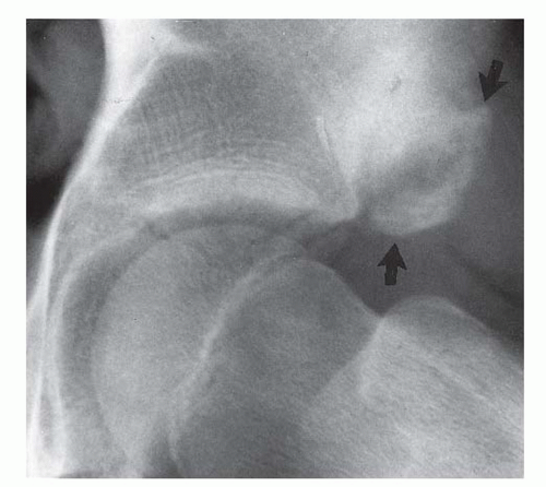
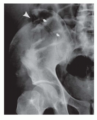
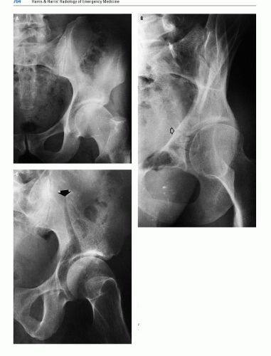
An inconsistent secondary ossification center may constitute the superior aspect of the posterior acetabular lip either unilaterally or bilaterally. These centers, which usually appear between the ages of 14 and 18 years and fuse in early adulthood, may persist ununited throughout adult life as the os acetabuli and simulate a posterior acetabular lip fracture (Fig. 17.11). As with ununited secondary ossification centers elsewhere, dense cortication of the margins of the separate center and smooth sclerotic contiguous surfaces of the adjacent portions of the pelvis and ununited center (Fig. 17.11) should easily distinguish these ununited secondary ossification centers from an acute fracture fragment.


Figure 17.8. Disparity in the radiographic appearance of the ischiopubic synchondroses (arrow) in an asymptomatic child. There was no history of antecedent trauma to the pelvis, and the bulbous configuration of the left ischiopubic synchondrosis is entirely normal.


Figure 17.9. Normal appearance of the inferior portion of the pelvis of a 14-year-old child. The triradiate cartilages (open arrow) are almost completely fused, whereas the ischiopubic synchondroses (black arrow) are fused.


Figure 17.10. Partially fused apophyseal centers of the iliac crest (arrows) and ischial tuberosity (arrowhead) (A) and of the pubic symphysis (arrows) (B).

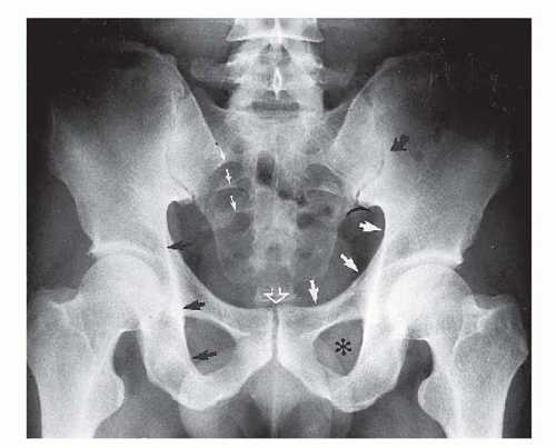
In the straight AP radiograph of the adult, the pelvic ring is the round or oval plane of the inlet of the true pelvis and includes the sacral promontory, the inferior margins of the SI joints, the iliopectineal line extending to the superior margin of the superior pubic rami, and the superior margin of the pubic symphysis. The pelvic ring is divided into an anterior and a posterior arch by an imaginary line connecting the ischial spines (Fig. 17.12). The ilioischial line (Figs. 17.1 and 
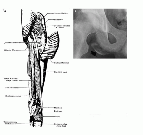
17.3) is not a single bony margin but is composed, superiorly, of the arc of the quadrilateral plate tangent to the x-ray beam and, inferiorly, by the internal cortex of the ischium extending to its tuberosity. The ilioischial line extends obliquely downward lateral to the iliopectineal line. The quadrilateral plate comprises the lateral surface of the true pelvis (birth canal) and, as such, constitutes the medial wall of the acetabulum.

The teardrop shadow of the pelvis (Fig. 17.13) is a composite U-shaped shadow located at the anteroinferior portion of the acetabular fossa and constitutes the anterior margin of the acetabular notch. It consists of cortical and medullary bone principally of the ischium with a small medial component from the superior pubic ramus.
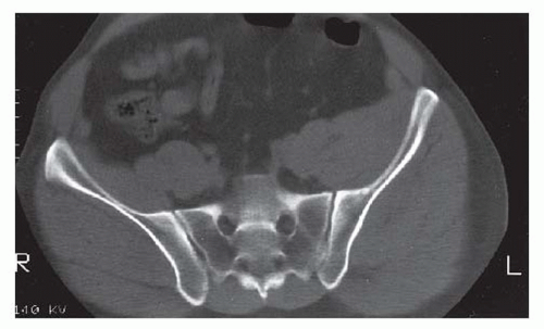
21


Figure 17.11. A: Frontal examination of the hips of a patient without symptoms referable to the pelvis. Open arrows indicate secondary ossification centers of the posterior lip of each acetabulum. B: The frontal projection of the hips of an adult demonstrates a persistent ununited secondary ossification center (os acetabuli) on the left. The radiographic characteristics of the contiguous surfaces of the os acetabuli and the posterior acetabular rim (arrow) should distinguish this normal variant from an acute posterior lip fracture.


Figure 17.12. Although not a component of the pelvic inlet, the ischial spines are the reference points used to divide the pelvic ring into its anterior and posterior arches.

The SI joints are oblique structures with the sacral alar component anterior to the iliac wing component. Consequently, the iliac and sacral alar margin of the anterior edge of the SI joint is usually clearly recognizable throughout its vertical extent. The posterior margin of the SI joint, which lies medial to the anterior margin, is much less well seen on the AP radiograph of the pelvis and is frequently identified only by the posterior margin of the iliac wing component (Fig. 17.3). Oblique views intended to demonstrate the SI joints en face usually only demonstrate the anterior aspect of the joint space. The SI joints are optimally demonstrated on axial CT images (Fig. 17.14). An important anatomic relationship is formed by the inferior cortical margins of the contiguous sacral alar and iliac surfaces of the anterior margins of the SI joints, which normally should be on the same plane or constitute a continuous imaginary arc (Fig. 17.1).


Figure 17.13. Pelvic “teardrop” (arrowheads).


Figure 17.14. Axial computed tomography of the normal sacrum and the SI joints of an adult.

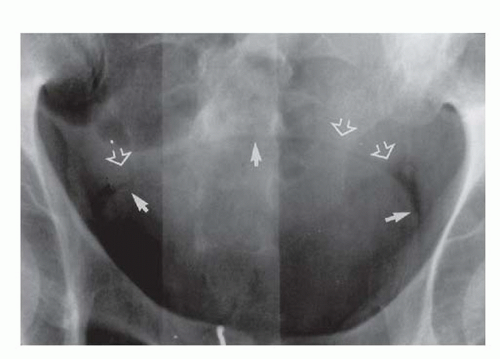
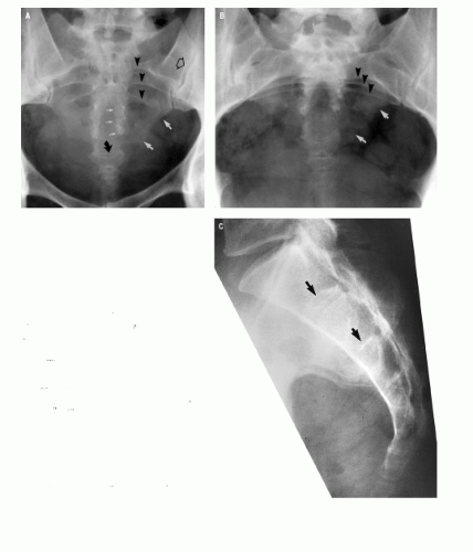
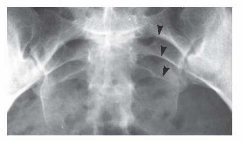
The sacral arcuate lines (Fig. 17.15), sometimes incorrectly referred to as “struts,” reflect the normal anatomy of the sacral foramina. The arcuate lines do not represent a specific cortical edge. Rather, they represent the arc of the superior surface of the sacral foramen that happens to be tangent to the x-ray beam. As described by Jackson et al., the sacral arcuate lines appear as rather sharply defined, superiorly convex curvilinear densities that are broad superomedially and taper gently and smoothly as they extend inferolaterally.22 The arcuate lines are normally equidistant from each other and are bilaterally symmetrical. Usually, the arcuate lines of only the upper three foramina are visible on the straight AP radiograph of the pelvis.


Figure 17.15. Normal sacral arcuate lines, sometimes improperly referred to as “struts” (arrowheads). These bilaterally symmetrical, arched, linear-appearing densities represent the arc of the sacral foramina tangent to the central x-ray beam.


RADIOGRAPHIC MANIFESTATIONS OF TRAUMA


Pelvic Injuries

Many classifications of pelvic fractures have been proposed. An early classification of pelvic fractures is Kane’s modification of the Key-Conwell classification (Table 17.1).23 A simplified and more inclusive version of the Kane classification of pelvic injuries prepared by the author is found in Table 17.2. The term “injuries” is preferred to “fractures” because some pelvic injuries are ligamentous disruptions and not fractures (e.g., pubic symphysis separation or SI joint diastasis).

The most widely accepted classification of PRD by orthopedic surgeons is that proposed by Pennal et al.24 and modified by Tile (Table 17.3).25, 26 More recently, Young et al.27 have proposed a classification of “pelvic injury” based on pattern of fragment distribution, similar to that of Pennal and Tile, that attempts to relate arterial bleeding to the fracture fragment pattern (Table 17.4).

The classifications of Kane, Pennal, and Tile are predicated on the mechanism of injury as deduced from the position of the fragments of the PRD on the initial AP radiograph (e.g., anteroposterior compression [APC], lateral compression [LC], and vertical shear [VS]). Pennal et al.24 noted, in proposing this basis for classification of pelvic fractures, that in fully one-third of the patients, the position of the fragments was consistent with more than one of the mechanisms of injury. Further, these classifications—either of pelvic fractures23 or PRD24, 25, 26, 27 —fail to take into account the work of Chenoweth et al.,28 Gertzbein and Chenoweth,29 and Bucholz,30 which conclusively demonstrate “that an injury at one site of the pelvic ring must be associated with another on the other side of the ring.”28 Furthermore, our own experience in the review of 300 initial AP radiographs of patients with PRD confirms that disruption of the anterior pelvic arch by either fracture or pubic symphysis diastasis must be associated with disruption of the posterior arch by either fracture or SI joint diastasis. Consequently, because of the animal and clinical research cited earlier, it is apparent that with the exception of the “insufficiency fracture,” stress fracture, or an anterior pelvic arch fracture in a child with unfused TRC, the type II fractures found in Kane’s modification of the Key-Conwell classification of pelvic fractures (Table 17.1) (e.g., “single breaks in the pelvic ring, occurring through both ipsilateral rami, one SI joint, or subluxation of the pubic symphysis”) simply do not exist following major blunt pelvic trauma.


TABLE 17.1 Kane”s Modification of Key-Conwell Classification of Pelvic Fractures


Type


Description


I


Breaks of individual bones not involving the pelvic ring. Includes avulsion fractures of a single ramus, and isolated fractures of the iliac wing, sacrum, or coccyx.


II


Single breaks in the pelvicring, occurring through both ipsilateral rami, one SI joint, or subluxatio of the symphysis pubis. By definition, there can be no displacement; otherwise, a second break in the ring must also be present.


III


Double breaks in the pelvic ring. Includes three subtypes:


1. Malgaigne variants, also called double vertical or dimetric fractures.


2. Bilateral double ramus fractures, referred to as either straddle fractures or butterfly pattern.


3. Severe multiple or crushing fractures.


IV


Acetabular fractures. Includes three subtypes:


1. Rim fractures.


2. Central acetabular fractures.


3. Ischioacetabular fractures.


From Kane WJ. Fractures of the pelvis. In: Rockwood CA, Green DP, eds. Fractures in Adults. 2nd ed. Philadelphia, PA: JB


TABLE 17.2 Classification of Pelvic Injuries (Harris)


I.


Injuries of isolated pelvic bones without disruption of pelvic ring


A.


Avulsion injuries


1. Anterosuperior iliac spine


2. Anterorinferior iliac spine


3. Ischial tuberosity apophysis


4. (Lesser trochanter apophysis, femur)


B.


Fractures of isolated bones, for example,


1. Iliac wing (Duverney), pelvic teardrop


2. Lower sacral segments


3. Coccyx


II.


Pelvic ring disruption


1.


Malgaigne—ipsilateral anterior and posterior arch disruption


2.


“Bucket-handle”—contralateral anterior and posterior arch disruption


3.


“Open-book”


4.


Other combinations of anterior and posterior arch disruption not identified by eponym


III.


Insufficiency fracture


IV.


Stress fracture


V.


Acetabular fracture


TABLE 17.3 Tile Classification of Pelvic Disruption


Type A


Stable


A1—Fractures of pelvis not involving the ring


A2—Stable, minimally displaced fractures of the ring


Type B


Rotationally unstable, vertically stable


B1—Open-book


B2—Lateral compression; ipsilateral


B3—Lateral compression; contralateral (bucket-handle)


Type C


Rotationally and vertically unstable


C1—Unilateral


C2—Bilateral


C3—Associated with an acetabular fracture


From Tile M. Pelvis ring fractures: should they be fixed?


J Bone Joint Surg Br. 1988;70:1, with permission.


TABLE 17.4 Injury Classification According to the Young System


Category


Distinguishing Characteristicsa


LC


Transverse fracture of pubic rami, ipsilateral, or contralateral to posterior injury


I—Sacral compression on side of impact


II—Crescent (iliac wing) fracture on side of impact


III—LC-I or LC-II injury on side of impact; contralateral open-book (APC) injury


APC


Symphyseal diastasis and/or longitudinal rami fractures


I—Slight widening of pubic symphysis and/or anterior SI joint; stretched but intact anterior SI, sacrotuberous, and sacrospinous ligaments; intact posterior SI ligaments


II—Widened anterior SI joint; disrupted anterior SI, sacrotuberous, and sacrospinous ligaments; intact posterior SI ligaments


III—Complete SI joint disruption with lateral displacement; disrupted anterior SI, sacrotuberous, and sacrospinous ligaments; disrupted posterior ligaments


VS


Symphyseal diastasis or vertical displacement anteriorly and posteriorly, usually through the SI joint, occasionally through the iliac wing and/or sacrum


CM


Combination of other injury patterns, with LC/VS being the most common


a “LC, lateral compression; APC, anteroposterior compression; SI, sacroiliac; VS, vertical shear; CM, combined mechanisms. From Young JW, Burgess AR, Brumback RJ, et al. Pelvic fractures: value of plain radiography in early assessment and management. Radiology. 1986;160(2):445-451 with permission.


Although the mechanism of injury classification of PRD has relevance in patient management, it is of little value in the identification of sites of PRD, which is the primary responsibility of the radiologist. Therefore, to facilitate recognition of PRD, we propose a classification of pelvic injuries that reflects the actual pathology of the injury and is based on the investigations of Chenoweth et al.,28 Gertzbein and Chenoweth,29 Bucholz,30 and our own experience. This classification of pelvic injuries, which includes PRD, appears in Table 17.2. Pelvic injuries are discussed in the sequence contained in Table 17.2.


Injuries of Isolated Pelvic Bones Without Disruptions of the Pelvic Ring


Avulsion Injuries

Avulsion injuries of pelvic apophyses are the result of sudden, maximum muscular effort, such as those requiring rapid acceleration (e.g., short-distance runners) or abrupt changes of speed or direction (e.g., football, basketball, soccer, lacrosse players). The athlete usually experiences a sudden sharp pain in the area of the avulsion and a “popping” or “snapping” sensation and is abruptly incapacitated, frequently falling.

Avulsion of the apophysis of the anterosuperior iliac spine (Fig. 17.16) is the result of violent contraction of the sartorius muscle. This apophysis is a thin piece of bone that is commonly difficult or impossible to see with routine pelvic radiographic technique. The history of the injury is so characteristic that if the avulsed anterior superior iliac spine apophysis is not visible on the routine AP pelvic radiograph, oblique views of the involved hemipelvis should be obtained with reduced (“soft tissue”) radiographic technique.


Figure 17.16. Avulsion of the apophysis of the anterosuperior iliac spine. The anatomic drawing (A) indicates the origin of the sartorius muscle from the anterior superior iliac spine. The radiograph (B) illustrates the common location of the thin, faintly dense, avulsed anterosuperior iliac spine apophysis (arrow).


The anteroinferior iliac spine apophysis is avulsed by the intact rectus femoris muscle (Fig. 17.17). This apophysis may be a large, obvious, separate piece of bone or a minimally displaced, small, and subtle fragment (Fig. 17.18). Rarely, both superior and inferior anterior iliac spine apophyses will be avulsed simultaneously (Fig. 17.19). The location and radiographic characteristics of a healed anteroinferior iliac spine avulsion (Fig. 17.20), coupled with a history of childhood “hip” injury, should distinguish this benign, posttraumatic finding from a primary bone neoplasm.

The hamstring muscles arise from the ischial tuberosity and in the adolescent athlete, violent contraction of these muscles results in avulsion of the ischial tuberosity apophysis (Fig. 17.21) rather than in the hamstring “pull” or “tear” commonly seen in more mature athletes. The older the adolescent and the larger the ischial tuberosity apophysis, the easier it will be to identify this injury (Fig. 17.21). When the avulsed apophysis is small and only faintly ossified, as occurs in younger children, the avulsed fragment may be difficult (Fig. 17.22A) or nearly impossible (Fig. 17.22B) to identify radiographically. In the latter circumstance, the diagnosis may be made by recognizing the subtle difference in the appearance of the injured and normal ischial tuberosity (Fig. 17.22B) coupled with the clinical correlation or by magnetic resonance imaging (MRI).


Figure 17.17. Avulsed anteroinferior iliac spine apophysis (arrows).


Figure 17.18. Minimally displaced avulsion of the anteroinferior iliac spine apophysis (arrow, A). The contralateral normal anterior iliac spine apophysis in this same patient is seen in B for comparison.


Figure 17.19. Simultaneous avulsion of the right superior (arrow) and inferior (arrowhead) iliac spine apophyses. Comparison of the inferior iliac spine apophyses in the AP radiograph of the pelvis (A) establishes the abnormal appearance on the right side. The avulsed anterosuperior spine apophysis is better demonstrated by using slightly underexposed (“soft tissue”) technique (B).

Avulsion of the apophysis of the lesser trochanter of the femur, although not an injury of the pelvis per se, is included here because of the similarity of the clinical presentation and the geographic proximity to the avulsive injuries of the pelvis. As the site of insertion of the iliopsoas muscle, the avulsed lesser trochanteric apophysis will be retracted proximally (Fig. 17.23).


Figure 17.20. Healed avulsion of the anteroinferior iliac spine apophysis (arrow).


Fractures of Isolated Bones of the Pelvis without Pelvic Disruption

The isolated iliac wing fracture (Duverney) that does not disrupt the integrity of the pelvic ring may be comminuted and readily detectable on the straight AP pelvic radiograph (Fig. 17.24) or may be so subtle as to require oblique projections (Fig. 17.25) for identification.

Isolated fractures of the lower sacral and the coccygeal segments are usually the result of a direct blow, as occurs in a fall when the patient lands in a sitting position. The sacral concavity precludes visualization of all sacral segments on a single straight AP radiograph of the pelvis. For adequate examination of the sacrum by plain radiography, frontal projections obtained with the x-ray tube angled cranially and caudally (Fig. 17.26) are required. These projections of the sacrum are very similar to those seen on the inlet and outlet views of the pelvis. However, isolated sacral (and coccygeal) fractures are best evaluated on the lateral projection of the sacrum and coccyx. Because the median sacral crest, which represents the fused sacral spinous processes, is extremely variable in appearance on the lateral radiograph, the detection of sacral fractures is best achieved by recognizing disruption of the anterior and posterior cortices of the fused sacral segments (Fig. 17.27). The presence of a presacral hematoma provides valuable supportive evidence of an acute sacral fracture.


Figure 17.21. A: Anatomic drawing of the muscles of the posterior aspect of the thigh, indicating the site of origin of the hamstring muscles from the ischial tuberosity. B: Radiographic appearance of avulsion of a mature iliac spine apophysis (arrow).


Figure 17.22. A: Partial avulsion (arrowheads) of an immature ischial tuberosity apophysis. B: The only indication that an extremely immature right ischial tuberosity apophysis has been avulsed is the irregularity of the surface of the right tuberosity (open arrows) compared with that of the normal left (arrows).


Figure 17.23. A: Schematic representation of avulsion of the apophysis of the lesser trochanter of the femur by the iliopsoas muscle. B: In the frontal radiograph, the rostrally retracted apophysis (arrow) is largely obscured by the femoral neck. C: With external rotation of the femur, the displaced apophysis (arrow) is clearly visible.


Figure 17.24. Severely comminuted, displaced fracture confined to the iliac wing (arrow and arrowheads) that does not disrupt the pelvic ring.


Figure 17.25. The iliac wing fracture line is barely perceptible in the routine frontal projection (A). Disruption of the iliopectineal line (open arrow) is visible in the anteriorly rotated oblique projection (B). Only in the posteriorly rotated oblique projection (C) is the extent of the fracture line and separation of the fragments (arrow) clearly visualized.


Figure 17.26. The cranially angled frontal projection of the sacrum (A) demonstrates the upper sacral segments very clearly, whereas the caudally angled frontal projection (B) demonstrates the lower sacral and coccygeal segments.


Figure 17.27. In the frontal projection of the sacrum (A), segments of the fracture line are indicated by the arrows. Because of the proximity of the fracture line to the intersegmental space, the fracture line could be interpreted as a portion of the intersegmental line. In the lateral projection (B), however, the fracture in the body of the fourth sacral segment is unambiguously evident (arrow).


The clinical diagnosis is much more reliable than the radiographic diagnosis of sacrococcygeal dislocation and coccygeal fracture. This is because of the normal great variability of the relationship of the coccyx to the last sacral segment and because of the size and configuration of the coccygeal segments.


Pelvic Ring Disruption

PRD is defined as interruption of the normal contour of the perimeter of the plane of the inlet of the true pelvis at two or more sites on opposite sides of the pelvic ring by either fracture or pubic symphyseal or SI joint diastasis. In adults, one or more sites of interruption must occur on opposite sides of the pelvic ring, commonly described as occurring in the anterior and posterior pelvic arches (Fig. 17.12).28, 29, 30 Two caveats pertain to this fundamental tenet. The first is that the pelvic ring may be disrupted at a single site in pediatric patients until fusion of the TRC (Fig. 17.28).31 The second relates to the coincidence of PRD and acetabular fractures. In our group of 300 patients with PRD, 41 (13.6%) sustained concurrent acetabular fractures. However, in no instance did the acetabular fracture constitute any component of the PRD. This seeming contradiction—that is, that acetabular fractures are not included as a component of PRD when, indeed, acetabular fractures do interrupt the continuity of the perimeter of the inlet to the true pelvis—is directly related to the definition of PRD, which requires interruption of the continuity of the pelvic ring at two or more sites on opposite sides of the ring. Acetabular fractures fail this definition by involving only one side of the pelvic ring.


Figure 17.28. The comminuted, slightly displaced fracture of the superior pubic ramus (arrow) is the only site of PRD in this 6-year-old child. The bulging ischiopubic synchondroses (arrowheads) are normal for a child of this age, and neither should be misinterpreted as a fracture site.

The concept of a concomitant PRD and acetabular fracture is illustrated in Figure 17.29. The PRD consists of the comminuted fracture of the left pubic body and inferior ramus coupled with the left sacral alar fracture. The minimally displaced transverse left acetabular fracture is clearly neither the anterior nor the posterior component of the PRD.

As noted earlier, many classifications of pelvic injuries and PRD have been proposed. With respect to PRD, the classifications of Tile26 (Table 17.3) and of Young et al.27, 32 are probably the most widely accepted. The Tile classification is based on stability or instability of the PRD, which in most instances is a clinical rather than a radiologic diagnosis. The Young classification is based on that of Pennal et al.24 and Tile and Pennal33 in which the mechanism of injury—for example, LC, APC, and VS—is determined by the position of the disrupted ring components on the initial AP radiograph. The Young (Table 17.4) classification further divides the LC and APC injuries into subgroups I, II, and III, depending on the degree of ring disruption. Because Pennal found that the pattern or distribution of disrupted ring components could be ascribed to two or more of the mechanisms of injury in approximately one-third of the patients, it seems reasonable that the same would apply to the Young classification. Although the Tile and Young classifications (Tables 17.3 and 17.4, respectively) are of unquestioned value in patient management decisions, neither directly addresses the primary responsibility of the radiologist in the emergency center, namely, the accurate diagnosis of PRD, which is based on detecting the site and type (e.g., fracture or joint diastasis) of the ring disruptions.

Prompted by the fact that 72% of sacral alar fractures were unrecognized by the faculty radiologists of our institution,22 which meant that PRDs were misdiagnosed in the same degree and stimulated by the work of Chenoweth et al.,28 the work of Gertzbein and Chenoweth,29 and the autopsy findings of Bucholz30 and frustrated by the radiologic diagnostic shortcomings of the Pennal and the Tile classifications of PRD, members of our department retrospectively reviewed the initial AP pelvic radiographs of 300 consecutive adult patients admitted to Hermann Hospital with PRD between January 1983 and December 1987 in the hope of developing a system of pattern recognition of PRD that would enhance and facilitate the plain radiograph diagnosis of PRD. The changes in contour, spatial relationships, and symmetry of the sacral arcuate lines caused by acute trauma (Fig. 17.30) as described by Jackson et al.22 were invaluable in the recognition of sacral alar fractures. Although we were well aware that the type of PRD (e.g., fracture or joint diastasis) is of great clinical significance, the type of ring disruption was considered immaterial for the purely diagnostic purposes of this study. Our method consisted of recording the sites of disruption on a schematic representation of the pelvic ring divided into anterior and posterior arches at the level of the ischial spines (Fig. 17.12). Ipsilateral superior and inferior pubic rami fractures were considered a single site of disruption, as was pubic diastasis with associated fracture. In the posterior pelvic arch, SI joint diastasis, fracture diastasis, or iliac or sacral alar fractures on the same side were considered a single site of ring disruption.

Only gold members can continue reading. Log In or Register to continue

Stay updated, free articles. Join our Telegram channel

Jun 20, 2016 | Posted by in GENERAL RADIOLOGY | Comments Off on Pelvis, Including Lower Urinary Tract Trauma

Full access? Get Clinical Tree

Get Clinical Tree app for offline access