(1)
Department of Radiology, Saitama Medical University, Moroyama, Saitama, Japan
Abstract
Its mean length is 38 mm and mean width is 13 mm. It becomes thinner as it approaches the tibia, and it is partially embedded within the joint capsule. Its tibial attachment site is located 1 cm below the articular surface.
4.1 Anatomy
PCL is also an intra-articular, extra-synovial structure.
Its mean length is 38 mm and mean width is 13 mm. It becomes thinner as it approaches the tibia, and it is partially embedded within the joint capsule. Its tibial attachment site is located 1 cm below the articular surface.
PCL is twice as thick as ACL, and it has twice as strong tensile strength as other ligaments in the knee. It receives abundant vascular supply.
PCL consists of two main fibrous bundles, that is, thicker anterolateral bundle and thinner posteromedial bundle. In normal condition, it is impossible to differentiate these two on MRI, and PCL appears as a one band with homogeneous texture. When PCL is damaged, only one of the two bundles may be torn (see later description).
PCL receives tension at all angles during flexion and extension of the knee.
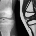
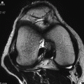
PCL runs within the intercondylar space in the superior-inferior direction, almost in parallel to the long axis of the knee (Fig. 4.1). It is therefore easy to visualize it on sagittal MRI. It is extremely rare not to be able to delineate PCL in a routine MRI examination.
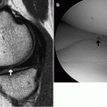
Meniscofemoral ligament is also called “the third cruciate ligament” and crosses in front of and behind PCL. This ligament connects the posterior horn of the lateral meniscus and the posterior surface of the medial femoral condyle. Note that it has the same name as the deep layer of the MCL (see later description). The portion of the meniscofemoral ligament that crosses in front of PCL is Humphrey’s ligament, and that crosses behind PCL is Wrisberg’s ligament. The latter is slightly larger than the former. Humphrey’s ligament is found in approximately 24% of patients, Wrisberg’s ligament in 23%, and both ligaments in 12% of patients (Fig. 4.2). These ligaments should not be mistaken as abnormality of PCL or menisci.
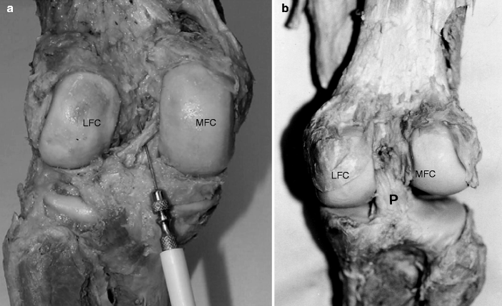
Fig. 4.1
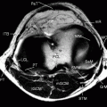
Cadaveric specimen showingWrisberg’s ligament (a) and PCL (b). Wrisberg’s ligament runs diagonally between the posterior horn of the lateral meniscus to the posterior surface of the medial femoral condyle (needle is inserted underneath the Wrisberg’s ligament, a). Deep to the Wrisberg’s ligament lies PCL, which runs vertically almost in parallel to the long axis of the knee (b). PCL is thick. LFC lateral femoral condyle, MFC medial femoral condyle, P PCL
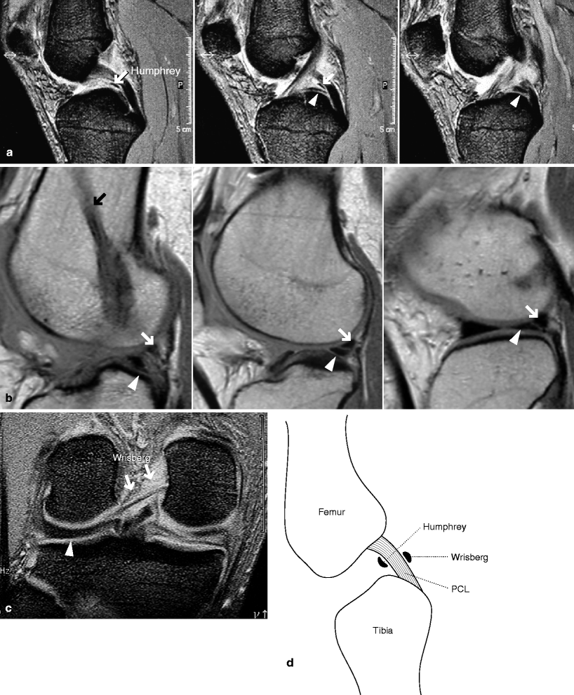
Fig. 4.2
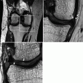
Humphrey’s and Wrisberg’s ligaments. Sagittal MRI demonstrates meniscofemoral ligament (Humphrey’s, white arrow in b, and Wrisberg’s, white arrow in (c). Coronal MRI also demonstrates Wrisberg’s ligament (c). These ligaments should not be mistaken as abnormalities of PCL or the lateral meniscus (arrowheads). The patient in (b) had ACL reconstruction (black arrow). (d) Schematic illustration showing the location of these ligaments
References
Sonin AH, Fitzgerald SW, Hoff FL, et al. MR imaging of the posterior cruciate ligament: normal, abnormal and associated injury patterns. Radiographics. 1995;15:551–61.
Grover JS, Bassett LW, Gross ML, et al. Posterior cruciate ligament: MR imaging. Radiology. 1990;174:527–30.
4.2 PCL Tear
PCL is thick and strong, and it is said to be less common than ACL tear (3–20% of all knee injuries, <1% of knee trauma requiring surgical intervention). However, this may be due to the fact that it is difficult to diagnose PCL tear clinically.
To have PCL torn, an enormous force is required, which is mostly due to traffic accident and rarely due to sports injury.
PCL injury alone is very rare, and often damages to ACL, collateral ligaments and menisci coexist.
Even if PCL is damaged and its function impaired or lost, the knee instability upon weight-bearing is less commonly seen than in ACL tear, and patients may not be aware of any symptoms. Damages to menisci and cartilage secondary to PCL tear is less commonly seen than in ACL tear.
Mechanism of injury for PCL tear
1.
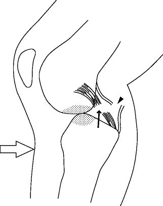
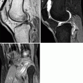
Direct force to the anterior aspect of the tibia: This is seen in “dashboard injury” in car accidents. Direct force is applied to the anterior aspect of the tibia when the knee is flexed, and the tibia is forcefully displaced posteriorly. This is the most common cause of PCL tear. In this mechanism, PCL is torn in the middle portion, and the posterior joint capsule may also be disrupted. Bone bruise may be seen in the posterior surface of the lateral femoral condyle and the anterior part of the tibial plateau (Fig. 4.3).

