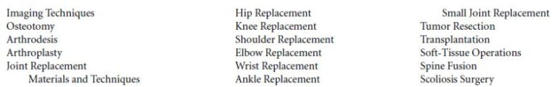
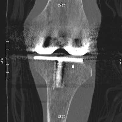
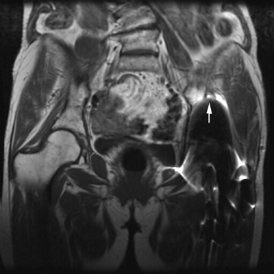
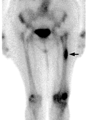
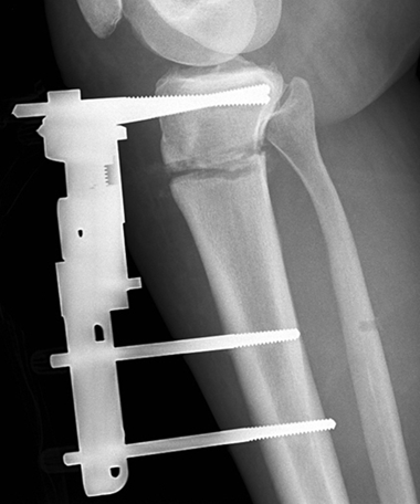
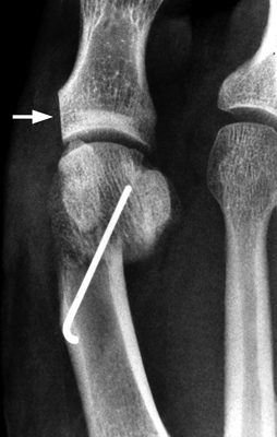
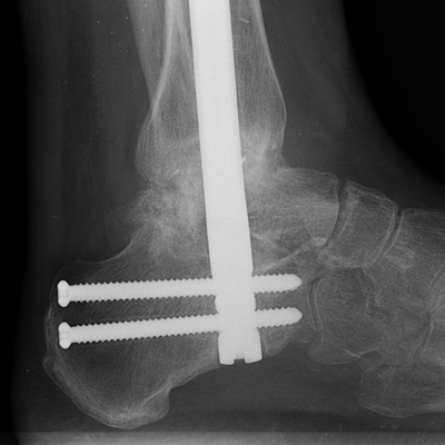
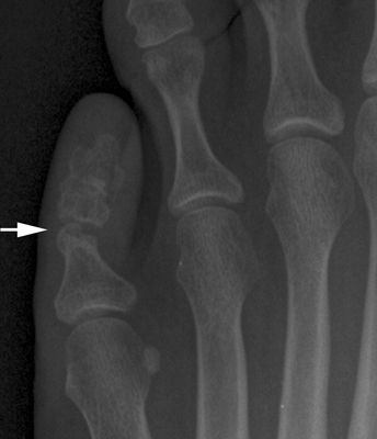
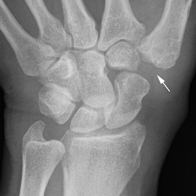
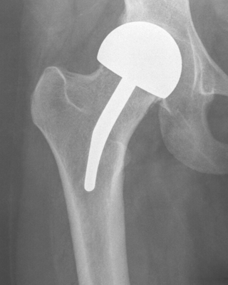
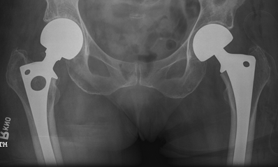
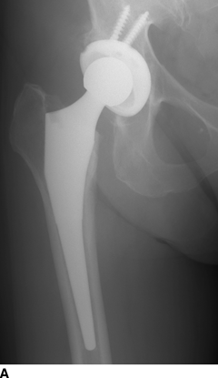
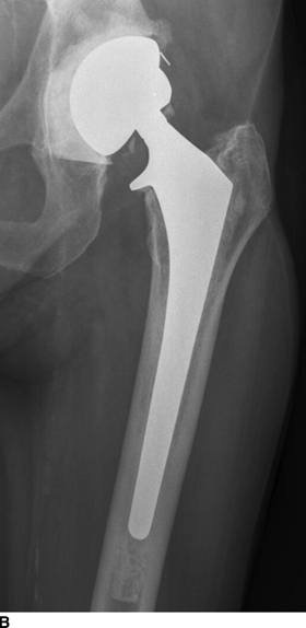
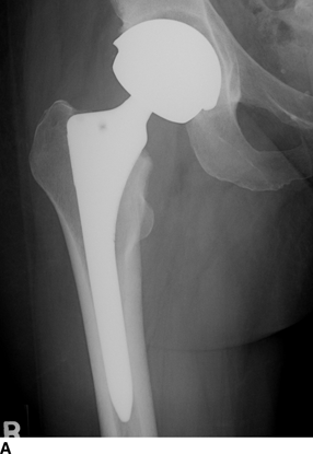
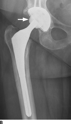
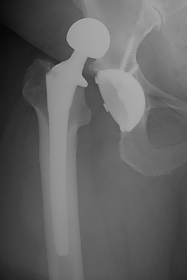
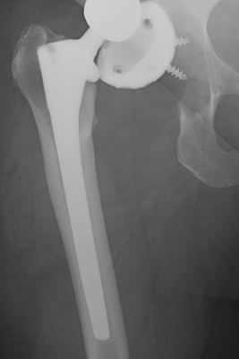
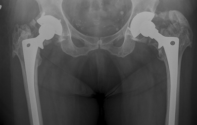
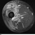
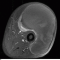
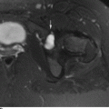
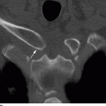
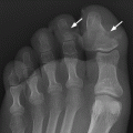
CHAPTER 17 Viewing images of postsurgical orthopedic patients can be mind-numbing if one does not understand the surgery that was performed. This chapter describes the imaging of the musculoskeletal system after a variety of orthopedic operations. Imaging after fracture treatment is described in Chapter 6. The standard diagnostic imaging techniques for the musculoskeletal system can be applied to postsurgical patients. Many operations are guided by the use of intraoperative fluoroscopy or radiographs. Standard radiographs are generally obtained immediately after the surgery to document the postoperative results. At follow-up visits, radiographs are obtained to monitor healing and to screen for complications. When extensive hardware has been implanted, nonstandard, obliquely positioned radiographs—or even radiographs positioned under fluoroscopy—may be necessary to project the hardware away from the bone, allowing it to be visualized. More specialized techniques are generally reserved for problem solving. CT can be used to evaluate the progress of healing when radiographs are equivocal, especially in the presence of complex anatomy and hardware. Streak artifacts on CT result from the high X-ray attenuation by metal (Fig. 17.1). Improvements in CT scanners and reconstruction algorithms and in orthopedic hardware materials have reduced the impact of these artifacts. On MRI, extensive artifacts may be caused by metallic implants (Fig. 17.2). Large implants may affect the surrounding magnetic field and cause the computer to distort the anatomy around the implants. Even when no hardware has been left behind, artifacts may be present from microscopic metallic particles introduced by instruments. These artifacts tend to be minimized on T1-weighted images but become more extensive (“bloom”) on T2-weighted or inversion recovery images (Fig. 17.3). However, previous surgery and orthopedic implants are not contraindications to MRI, and MRI may provide useful information in many situations. Arthrography should be considered when there is a question of joint infection or other postsurgical joint abnormalities. Stress views or examination under fluoroscopy may document motion at sites where none was intended. Radionuclide bone scan is useless in the immediate postoperative period because of tracer uptake at the surgical site. However, tracer accumulation returns toward normal after approximately 6 months, after which time the bone scan may be helpful (Fig. 17.4). Imaging with labeled leukocytes or gallium-67 may be indicated when there is a question of infection. FIGURE 17.1. Coronal reconstruction of CT scan through TKR shows severe artifact from the presence of metal, yet the metal-bone interface (arrow) of the tibial component is clearly visible. FIGURE 17.2. Coronal T2-weighted MRI of the pelvis with major metal artifact on the left side due to the presence of a total hip replacement. The artifact (arrow) extends well above the actual location of the replacement. FIGURE 17.3. MR with metal artifact from metal surgical instruments. T1-weighted axial spine MRI shows very dark areas (arrow) overlying the skin and the left posterolateral spinal canal in this patient with laminectomy. There were no actual metal clips or other implants present. FIGURE 17.4. Anterior view of the radionuclide bone scan of the patient with bilateral total hip replacements. A stress fracture was present in the lateral femoral cortex on the left (arrow). An osteotomy is any operation that involves a surgical cut through the bone. Osteotomies are commonly performed to change the alignment, length, or shape of a bone. For example, a fracture malunion with angular deformity may be revised by an osteotomy that realigns the fragments. Osteotomies may be described by their site and the direction of change in the distal fragment. A valgus osteotomy of a long bone realigns the distal fragment into the valgus relative to its original alignment. A lateral displacement osteotomy repositions the distal fragment laterally. Changes in the rotational alignment may also be made; these are called rotational osteotomies. Shortening is accomplished by the removal of a bone. Lengthening may be accomplished by displacement of overlapping fragments by the insertion of a bone graft or by bone distraction using an external fixator. The fixation and healing of osteotomies are similar to that of fractures, and the hardware devices used to treat fractures are applicable (see Chapter 6). Complications of healing are rare. Osteotomies are also used to alter the alignment and biomechanics of joints. A high tibial valgus osteotomy is a common treatment for severe medial compartment osteoarthritis with varus deformity (Fig. 17.5). By realigning the tibial shaft, the varus deformity is corrected, and the mechanical axis of the knee is restored. This has the effect of reducing the weight-bearing stress on the diseased medial compartment and redistributing some of it to the lateral compartment. Distal femoral varus osteotomies can correct valgus deformities at the knee from the lateral compartment osteoarthritis. Similarly, angular or rotational osteotomies of the proximal femur alter the major weight-bearing portion of the femoral head in the treatment of hip dysplasia, early osteoarthritis, or similar conditions. An innominate osteotomy may be used to deepen the acetabulum. FIGURE 17.5. High tibial valgus osteotomy with external fixator. A bunion is a symptomatic protrusion at the medial aspect of the head of the first metatarsal that is the result of a valgus deformity of the great toe and a varus deformity of the first metatarsal (also called hallux valgus primus varus). Tension along the flexors and extensors of the great toe acts like bowstrings to increase the angular deformities. The condition may be painful and is often compounded by the difficulty of fitting shoes. Surgical correction involves not only removing the bony prominence but also—more important—realigning the great toe with its flexors and extensors. Commonly, the first metatarsal is shortened, and the head is displaced laterally (Fig. 17.6). FIGURE 17.6. Bunion repair with shortening and displacement osteotomy of the first metatarsal, fixed by K-wire. Also note osteotomy of the proximal phalanx of the great toe (arrow). An arthrodesis is a surgical fusion of a joint. An intra-articular arthrodesis is accomplished by resecting the joint and fixing the ends of the articulating bones together. Internal or external fixation may be used as well as bone graft. A healed and remodeled arthrodesis has a continuous cortex and trabecular structure across the site. Adaptive changes may occur in adjacent joints in response to the loss of motion. Joint pain in arthritis can be eliminated by synovectomy and arthrodesis. Common sites for arthrodesis include the fingers, for treatment of arthritis; the wrist, for treatment of arthritis or posttraumatic instability; and the subtalar joint, for treatment of posttraumatic arthritis. One of the more common arthrodeses involving the foot is the triple arthrodesis, in which the subtalar joints, calcaneocuboid joints, and talonavicular joints are arthrodesed (Fig. 17.7). In the setting of arthritis, it may sometimes be difficult to distinguish an arthrodesis from a bony ankylosis (joint fusion caused by a disease). An extra-articular arthrodesis is accomplished by establishing a bony bridge across a joint that prevents motion without actually resecting the joint itself. Some spine fusions involve an extra-articular arthrodesis. An epiphysiodesis, a surgical fusion of an open growth plate, may be performed for correction of growth discrepancy. FIGURE 17.7. Ankle arthrodesis with locked IM rod fixation. An arthroplasty is a surgical repair of a joint. Resection arthroplasty involves excision of one or both of the articular surfaces, leaving the ends of the bone to articulate with each other. Because the joint is not fixed, a pseudoarthrosis develops, preserving motion. Common sites for resection arthroplasty include the toes and hip. Many toe deformities result from soft-tissue contractures. Reducing the length of the bone through resection arthroplasty eliminates the effect of contractures (Fig. 17.8). Resection arthroplasty may be performed with resection of a small bone at a painful joint such as the trapezium from the first carpometacarpal (CMC) joint (Fig. 17.9). At the hip, a resection arthroplasty is called a Girdlestone procedure. Replacement arthroplasty involves replacing one or more articular surfaces with a prosthesis (discussed in “Joint Replacement”). FIGURE 17.8. Resection arthroplasty of the IP joint (arrow) of the fifth toe. FIGURE 17.9. Resection arthroplasty of the first CMC joint (arrow). Prosthetic joint replacements are the most common elective orthopedic operations performed in the United States. A total joint replacement arthroplasty replaces both sides of the joint with prosthetic components. A hemiarthroplasty replaces one side of the joint with a prosthesis. Metal, polyethylene, and cement are commonly used in manufacturing joint replacements. Metal components—usually titanium or a cobalt-chromium alloy—are used for strength and stiffness. The fatigue strength of these metals under cyclic loading is such that in vivo failure is rare. Ultrahigh-molecular-weight polyethylene is used for concave articular surfaces such as the acetabulum, tibial plateau, and glenoid fossa. This material is denser and stiffer than the polyethylene used in common kitchenware and is manufactured to have high resistance to abrasive wear. A metal back is often used to provide mechanical support. Polyethylene is radiolucent—like other plastics—but the presence of a metal back, especially at the acetabulum, may obscure the polyethylene on radiographs. Many polyethylene components have embedded metal markers that indicate their position on radiographs. Bone cement (polymethyl methacrylate or methyl methacrylate) is a rapidly polymerizing acrylic plastic that is used as an adhesive to fix metallic or polyethylene components to the bone. Cement is rendered radiopaque during manufacture by the addition of barium sulfate. Ceramics have become more popular for use as articular surfaces. For the small joints of the hands and feet, silicone rubber prostheses may be used. Recently, pyrolytic carbon components have been introduced for use in the hand. Implanted metal components are often fixed into the bone with cement. Bubbles in the cement that might act as stress risers are removed before use by centrifugation or vacuum chamber, and the cement is injected into the medullary space under high pressure. A polyethylene plug at the bottom of the prepared cavity creates a closed space and prevents flow of cement down the medullary canal. Metal prostheses that are implanted without cement may have a mechanical press fit that provides immediate stability and a specially textured surface (porous coat) that allows ingrowth of the bone or fibrous tissue, providing eventual long-term fixation. A high degree of precision is ensured at surgery through the use of jigs, cutting guides, and mock-ups that allow the surgeon to achieve a perfect geometric fit between the bone and the prosthetic component. Screws temporarily secure some cementless components until bone ingrowth occurs. Hydroxyapatite crystals applied to the surfaces of cementless metal components during manufacturing may improve biologic fixation in the bone. The crystalline coating of the prosthesis is directly incorporated into the molecular structure of the host bone. Because different materials deform to different degrees under mechanical loading (their stiffness is different), shear stresses develop at the interfaces, particularly between the bone and metal or the bone and cement. A layer of fibrous tissue that grows into these interfaces helps dissipate the forces by redistributing them over a greater surface area. This layer is similar to the periodontal ligament that cushions the teeth in the softer cancellous bone of the jaws. The fibrous layer may be visible on radiographs as a fine lucency (less than 2 mm thick) between the bone and cement or the bone and metal. An additional area of increased stress is the interface between the cement and metal; microscopic motion may occur at this interface, but no gap should be seen. There are several general types of hip replacements. A cup arthroplasty resurfaces the articular surface of the femoral head with a metal cup (Fig. 17.10). Hemiarthroplasties of the hip replace the femoral head and neck (Fig. 17.11). Simple femoral prostheses compose a single metal component with a stem that is fitted into the medullary canal and a head that articulates with the native acetabulum. Bipolar femoral prostheses have a metal femoral component comprising a stem and a head and an acetabular component comprising a metal socket with a polyethylene liner. The metal socket articulates with the native acetabulum, and the head of the femoral component articulates with the polyethylene liner. Although the acetabular component is not fixed, most of the motion takes place between the head and liner, preserving the native acetabular cartilage. Hemiarthroplasties are used for the disease of the proximal femur in which the acetabulum is relatively normal. For example, a femoral neck fracture complicated by osteonecrosis might be managed with a bipolar femoral prosthesis. A bipolar prosthesis may be converted to a total hip replacement (THR) with interchangeable components. A THR has both femoral and acetabular components that replace the native articular surfaces (Fig. 17.12). Most currently implanted prostheses have a metal acetabular component with a polyethylene liner that forms the articular surface, so that the bearing surface is metal-on-polyethylene. Some newer implants have metal-on-metal, ceramic-on-polyethylene, or ceramic-on-ceramic bearing surfaces (Fig. 17.13). The components may be fixed to the bone with or without cement. Some surgical approaches require an osteotomy of the greater trochanter for exposure; it is usually reattached with wires or cables. If a nonunion develops, a lurching gait disturbance results. If a gluteal soft-tissue release is used for surgical exposure, the gluteal musculature may be reattached to the greater trochanter with sutures, screws with washers, or soft-tissue anchors. Bone graft and specialized hardware may be inserted to buttress a deficient proximal femur or acetabulum. FIGURE 17.10. Resurfacing hemiarthroplasty of the hip. FIGURE 17.11. Bilateral femoral endoprostheses, unipolar on the right and bipolar on the left. FIGURE 17.12. Total hip replacements (different patients). A: Noncemented THR. B: Cemented THR. FIGURE 17.13. Total hip replacements (different patients). A: Noncemented THR with metal-on-metal bearing surface. B: Noncemented THR with ceramic head (arrow). The most common early complication of THR is dislocation. Typically, the femoral head rotates externally out of the acetabulum (Fig. 17.14). Loss of fixation of the acetabular cup may allow it to dislocate from the acetabular bed (Fig. 17.15). FIGURE 17.14. Dislocated THR. The head has moved laterally and superiorly. FIGURE 17.15. Dislocation of the acetabular cup from the acetabular bed, with dislocation of the femoral head from the cup. Heterotopic bone formation after THR is common; it may occasionally interfere with motion (Fig. 17.16). FIGURE 17.16. Heterotopic ossification in bilateral THRs.
Postsurgical Imaging
 IMAGING TECHNIQUES
 OSTEOTOMY
 ARTHRODESIS
 ARTHROPLASTY
 JOINT REPLACEMENT
Materials and Techniques
Hip Replacement
Related posts:
Stay updated, free articles. Join our Telegram channel

Full access? Get Clinical Tree


