Radiographic Exposure Technique


Radiographic Exposure Technique


Introduction


In Chapter 6, variables that affect both the quantity and quality of the x-ray beam are presented. Milliamperage and time affect the quantity of radiation produced and kilovoltage affects both the quantity and quality. Chapter 9 emphasizes that a good-quality radiographic image accurately represents the anatomic area of interest. The characteristics evaluated for image quality are density or brightness, contrast, recorded detail or spatial resolution, distortion, and noise. This chapter focuses on radiographic exposure techniques and the use of accessory devices, and their effect on the radiation reaching the image receptor (IR) and the image produced. Radiographers have the responsibility of selecting the combination of exposure factors to produce a quality image. Knowledge of how these factors affect the image individually and in combination assists the radiographer to produce a radiographic image with the amount of information desired for diagnostic interpretation.


Because various types of IRs respond differently to the radiation exiting the patient (remnant), these differences are noted throughout this chapter. However, regardless of the type of IR, the patient should be exposed to the least amount of radiation necessary to produce a diagnostic-quality radiograph. This chapter discusses all of the primary and secondary factors and their effects on the radiation reaching the IR.


Primary Factors


The primary exposure technique factors the radiographer selects on the control panel are milliamperage, time of exposure, and kilovoltage peak (kVp). Depending on the type of control panel, milliamperage and exposure time may be selected separately or combined as one factor, milliamperage/second (mAs). Regardless, it is important to understand how changing each separately or in combination affects the radiation reaching the IR and the radiographic image.


Milliamperage and Exposure Time


The quantity of radiation reaching the patient affects the amount of remnant radiation reaching the IR. The product of milliamperage and exposure time has a direct proportional relationship with the quantity of x-rays produced.


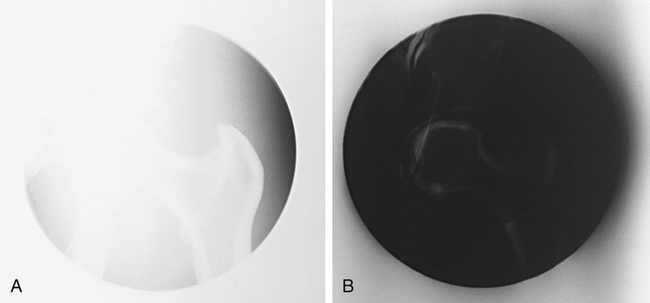
Once the anatomic part is adequately penetrated, as the quantity of x-rays is increased, the exposure to the IR proportionally increases (Figure 10-1). Conversely, when the quantity of x-rays is decreased, the exposure to the IR decreases. Therefore exposure to the IR can be increased or decreased by adjusting the amount of radiation (mAs).


Because the mAs is the product of milliamperage and exposure time, increasing milliamperage or time has the same effect on the radiation exposure.


As demonstrated in Math Application 10-1, mAs can be doubled by doubling the milliamperage or doubling the exposure time. A change in either milliamperage or exposure time proportionally changes the mAs. To maintain the same mAs, the radiographer must increase the milliamperage and proportionally decrease the exposure time.


It is important for the radiographer to determine the amount of mAs needed to produce a diagnostic image. This is not an easy task because there are so many variables that can affect the amount of mAs required. For example, single-phase generators produce less radiation for the same mAs when compared with a high-frequency generator.


A patient’s age, condition, and the presence of a pathologic condition also affect the amount of mAs required for the procedure. Additionally, for a given mAs, IRs respond differently. For film-screen IRs, the mAs controls the density produced in the image. There is a direct relationship between the amount of mAs and the amount of density produced when using film-screen IRs. For example, when the mAs is increased, density is increased; when the mAs is decreased, density is decreased (Figure 10-2).


When a film image is too light (insufficient density), a greater increase in mAs may be needed to correct the density, or the mAs may need to be decreased to correct a film image that has excessive density. This relationship between radiation exposure intensity and density is discussed in more detail in Chapter 9. The film characteristic, speed, and chemical processing determine the amount of optical density produced on the image for a given mAs.


When using a film-screen IR, radiographers need to assess the level of density produced on the processed image and determine whether the density is sufficient to visualize the anatomic area of interest. When the radiograph is deemed unacceptable, this means the optical densities lie outside the film’s sensitometric curve’s straight-line portion, and may need to be repeated. The radiographer must decide how much of a change in mAs is needed to correct for the density error.


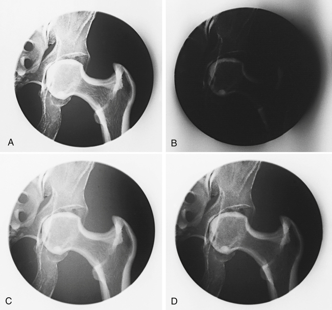
In general, for repeat radiographs necessitated by density errors, the mAs is adjusted by a factor of 2; therefore a minimum change involves doubling or halving the mAs. This typically brings the optical densities back within the straight-line portion of the film’s sensitometric curve to best visualize the anatomic area of interest. As mentioned previously, it may take more than doubling the mAs to correct for a density error. If the radiograph necessitates an adjustment greater than a factor of 2, the radiographer should multiply or divide the mAs by 4 (Figure 10-3).


Radiographs that have sufficient but not optimal density usually are not repeated. If a radiograph must be repeated because of another error, such as positioning, the radiographer may also use the opportunity to make an adjustment in density to produce a radiograph of optimal quality. Making a visible change in radiographic density requires that the minimum amount of change in mAs be approximately 30% (depending on equipment, this may vary between 25% and 35%). Radiographic images generally are not repeated to make only a slight visible change. A radiographic image repeated because of insufficient or excessive density requires a change in mAs by a factor of at least 2.


Digital IRs can detect a wider range of radiation intensities (wider dynamic range) exiting the patient and therefore are not as dependent on the mAs as film-screen IRs. However, exposure errors can adversely affect the quality of the digital image. If the mAs is too low (low exposure to the digital IR), image brightness is adjusted during computer processing to achieve the desired level. Although the level of brightness has been adjusted, there may be increased quantum noise visible within the image. If the mAs selected is too high (high exposure to the digital IR), the brightness can also be adjusted, but the patient has received more radiation than necessary.


The radiographer should be diligent in monitoring exposure indicator values to ensure that quality images are obtained with the lowest possible radiation dose to the patient.


To best visualize the anatomic area of interest, the mAs selected must produce a sufficient amount of radiation reaching the IR, regardless of type. Excessive or insufficient mAs adversely affects image quality and affects patient radiation exposure.


Kilovoltage


The kVp affects the exposure to the IR because it alters the amount and penetrating ability of the x-ray beam.


The area of interest must be adequately penetrated before the mAs can be adjusted to produce a quality radiographic image. When adequate penetration is achieved, further increasing the kVp results in more radiation reaching the IR. Unlike mAs, the kVp affects the amount of radiation exposure to the IR and radiographic contrast.


However, the kVp has a greater effect on the image when using film-screen IRs. Increasing the kVp increases IR exposure and the density produced on a film image, and decreasing the kVp decreases IR exposure and the density produced on a film image (Figure 10-4).


For film-screen IRs, kVp has a direct relationship with density; however, the effect of the kVp on density is not equal throughout the range of kVp (low, middle, and high). A greater change in the kVp is needed when operating at a high kVp (greater than 90) compared with operating at a low kVp (less than 70) (Figure 10-5).


Because kVp affects the amount of radiation reaching the IR, its effect on the digital image is similar to the effect of mAs. Too much radiation reaching the IR (within reason) produces a digital image with the appropriate level of brightness as a result of computer adjustment during image processing; however, the patient has been overexposed. Similarly, too little radiation reaching the IR (within reason) produces a digital image with the appropriate level of brightness, but the increased noise decreases image quality.


Kilovoltage is not a factor typically manipulated to vary the amount of IR exposure in film-screen imaging because the kVp also affects contrast. However, it is sometimes necessary to manipulate the kVp to maintain the required exposure to the IR. For example, using portable or mobile x-ray equipment may limit choices of mAs settings and therefore the radiographer must adjust the kVp to maintain sufficient exposure to the IR.


Maintaining or adjusting exposure to the IR can be accomplished with kVp by using the 15% rule. The 15% rule states that changing the kVp by 15% has the same effect as doubling the mAs, or reducing the mAs by 50%; for example, increasing the kVp from 82 to 94 (15%) produces the same exposure to the IR as increasing the mAs from 10 to 20.


Increasing the kVp by 15% increases the exposure to the IR, unless the mAs is decreased. Also, decreasing the kVp by 15% decreases the exposure to the IR, unless the mAs is increased. As mentioned earlier, the effects of changes in the kVp are not uniform throughout the range of kVp. When low or high kVps are used, the amount of change in the kVp required to maintain the exposure to the IR may be greater or less than 15%.


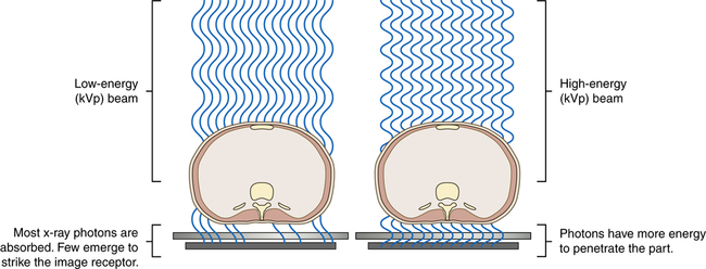
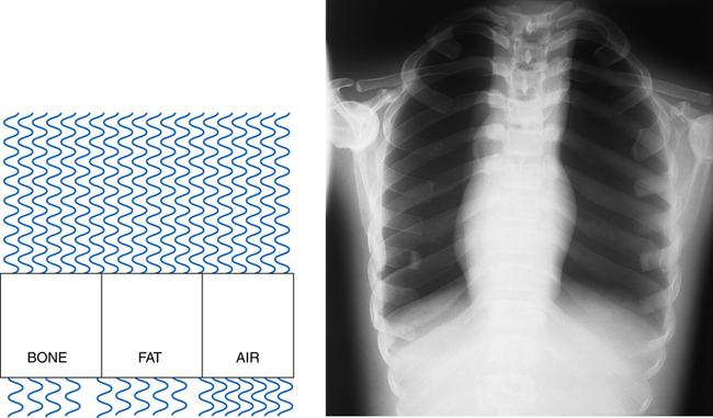
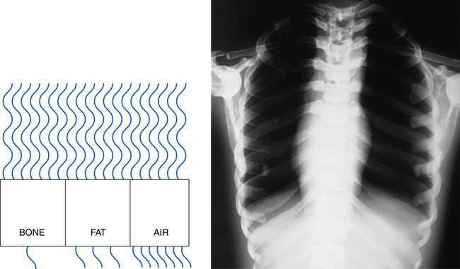
Altering the penetrating power of the x-ray beam affects its absorption and transmission through the anatomic tissue being radiographed. Higher kVp increases the penetrating power of the x-ray beam and results in less absorption and more transmission in the anatomic tissues, which results in less variation in the x-ray intensities exiting the patient (remnant). As a result, images with lower contrast are produced (Figure 10-6). When a low kVp is used, the x-ray beam penetration is decreased, resulting in more absorption and less transmission, which results in greater variation in the x-ray intensities exiting the patient (remnant). This produces a high-contrast radiographic image (Figure 10-7).


Changing the kVp affects its absorption and transmission as it interacts with anatomic tissue; however, using a higher kVp reduces the total number of interactions and increases the amount of x-rays transmitted. In these interactions, more Compton scattering than x-ray absorption occurs (photoelectric effect) and more scatter exits the patient.


The level of radiographic contrast desired, and therefore the kVp selected, depends on the type and composition of the anatomic tissue, the structures that must be visualized, and to some extent the diagnostician’s preference. These factors make achieving a desired level of radiographic contrast more complex than achieving a desired level of radiographic density, especially for film-screen imaging.


For most anatomic regions, an accepted range of kVp provides an appropriate level of radiographic contrast. As long as the kVp selected is sufficient to penetrate the anatomic part, the kVp can be further manipulated to alter the radiographic contrast.


Radiographs generally are not repeated because of contrast errors. More often, the radiographer evaluates the level of contrast achieved to improve the contrast for additional radiographs or similar circumstances that arise with a different patient. If a repeat radiograph is necessary and kVp is to be adjusted to either increase or decrease the level of contrast, the 15% rule provides an acceptable method of adjustment. In addition, whenever a 15% change is made in the kVp to maintain the exposure to the IR, the radiographer must adjust the mAs by a factor of 2. Remember that a 15% change in kVp does not produce the same effect across the entire range of kVp used in radiography. A greater increase is needed for high kVp (90 and above) than for low kVp (below 70).


Adequate penetration of the anatomic area of interest is equally important when using digital IRs, and therefore kVp selection is important in producing a quality image. Assuming that the anatomic part is adequately penetrated, changing the kVp does not affect the digital image in the same way as a film-screen image. The kVp affects the contrast in a digital image; however, image brightness and contrast are primarily controlled during computer processing. When a kVp that is too low is selected, the brightness and contrast are adjusted, but quantum noise may be visible. Additionally, when a kVp that is too high is selected, the image brightness and contrast are adjusted, but patient exposure may be increased. Although image contrast can be adjusted when using a kVp that is too high, increased scatter radiation reaches the IR and may adversely affect image quality.


The selection of kVp alters its absorption and transmission through the anatomic part regardless of the type of IR used and therefore must be selected wisely. Exposure techniques using higher kVp with lower mAs exposure techniques are recommended in digital imaging because contrast is primarily controlled during computer processing. Higher kVp and lower mAs values are not recommended as a general rule during film-screen imaging because of the contrast required to best visualize the anatomic structures.


Secondary Factors


Many secondary factors affect the radiation reaching the IR and image quality. It is important for the radiographer to understand their effects individually and in combination.


Focal Spot Size


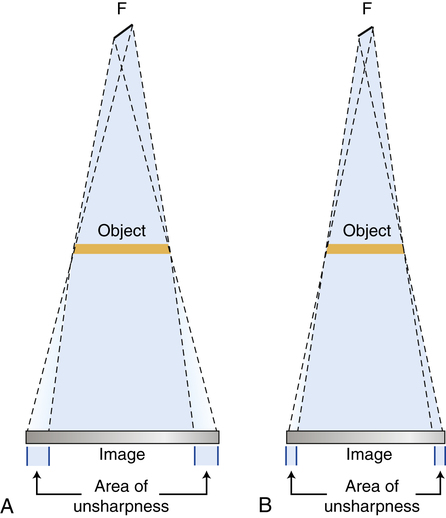
On the control panel the radiographer can select whether to use a small or large focal spot size. The physical dimensions of the focal spot on the anode target in x-ray tubes used in standard radiographic applications usually range from 0.5 to 1.2 mm. Small focal spot sizes are usually 0.5 or 0.6 mm, and large focal spot sizes are usually 1 or 1.2 mm in size. Focal spot size is determined by the filament size. When the radiographer selects a particular focal spot size, he or she is actually selecting a filament size that is energized during x-ray production. Focal spot size is an important consideration for the radiographer because the focal spot size affects recorded detail.


In general, the smallest focal spot size available should be used for every exposure. Unfortunately, exposure is limited with a small focal spot size. When a small focal spot is used, the heat created during the x-ray exposure is concentrated in a smaller area and could cause tube damage. The radiographer must weigh the importance of improved recorded detail for a particular examination or anatomic part against the amount of radiation exposure used. Modern radiographic x-ray generators are equipped with safety circuits that prevent an exposure from being made if that exposure exceeds the tube loading capacity for the focal spot size selected. Repeated exposures made just under the limit over a long period can still jeopardize the life of the x-ray tube.


Feb 27, 2016 | Posted by in GENERAL RADIOLOGY | Comments Off on Radiographic Exposure Technique

Full access? Get Clinical Tree

Get Clinical Tree app for offline access