Radiologic Evaluation of Tumors and Tumor-like Lesions


Radiologic Evaluation of Tumors and Tumor-like Lesions


Classification of Tumors and Tumor-like Lesions

Tumors, including tumor-like lesions, can generally be divided into two groups: benign and malignant. The latter group can be further sub-classified into primary malignant tumors, secondary malignant tumors (from the transformation of benign conditions), and metastatic tumors (Fig. 16.1). All of these lesions can be still further classified according to their tissue of origin (Table 16.1). Table 16.2 lists benign conditions that have the potential for malignant transformation.

To understand the terminology applied to tumors and tumor-like lesions of the bone, it is important to redefine certain terms pertinent to lesions and their location in the bone. The term tumor generally means mass; in common radiologic and orthopedic parlance, however, it is the equivalent of the term neoplasm. By definition, a neoplasm, ruled by an uncontrolled process of aberrant cellular and morphologic mechanisms, demonstrates autonomous growth; if in addition it produces local or remote metastases, it is defined as a malignant neoplasm or malignant tumor. Beyond this (and not dealt with in this chapter) are specific histopathologic criteria for defining a tumor as benign or malignant. It is nevertheless worth mentioning that certain giant cell tumors, despite a “benign” histopathology, may produce distant metastases, and that certain cartilage tumors, despite adhering to a “benign” histopathologic pattern, can behave locally like malignant neoplasms, even though this is detectable only radiologically. Moreover, certain lesions discussed here and termed tumor-like lesions are not true neoplasms, but rather have a developmental or inflammatory origin. They are included in this chapter because they display an imaging pattern that is almost indistinguishable from that of true neoplasms. Their cause is, in some cases, still being debated.

Equally important is the redefinition of certain terms pertinent to the location of a lesion in the bone. In the growing skeleton, one can clearly distinguish the epiphysis, growth plate, metaphysis, and diaphysis (Fig. 16.2A), and when lesions are located at these sites they are named accordingly. The greatest confusion is in the use of the term metaphysis. The metaphysis is a histologically very thin zone of active bone growth, adjacent to the physis (growth plate). Consequently, for a lesion to be called metaphyseal in location, it must extend into and abut the growth plate. However, it is customary—however incorrect—to use the same term for locating a lesion after skeletal maturity has occurred. By the time of maturity, the growth plate is scarred, and neither the epiphysis nor metaphysis remains. More proper and less confusing would be a terminology (Fig. 16.2B) such as articular end of the bone and shaft for locating lesions in the bone whose growth plate has been obliterated and whose metaphysis has ceased to exist. Some other terms used to describe the location of bone lesions are illustrated in Figure 16.3.


Radiologic Imaging Modalities

In general, the imaging of musculoskeletal neoplasms can be considered from three standpoints: detection, diagnosis (and differential diagnosis), and staging (Fig. 16.4). The detection of a bone or a soft-tissue tumor does not always require the expertise of a radiologist. The clinical history and the physical examination are often sufficient to raise the suspicion of a tumor, although radiologic imaging is the most common means of revealing one. The radiologic modalities most often used in analyzing tumors and tumor-like lesions include (a) conventional radiography; (b) angiography (usually arteriography); (c) computed tomography (CT); (d) magnetic resonance imaging (MRI); (e) scintigraphy (radionuclide bone scan); (f) positron emission tomography (PET) and PET-CT; and (g) fluoroscopy-guided or CT-guided percutaneous soft-tissue and bone biopsy.


Conventional Radiography

In most instances, the standard radiographic views specific for the anatomic site under investigation suffice to make a correct diagnosis (Fig. 16.5), which can subsequently be confirmed by biopsy and histopathologic examination. Conventional radiography yields the most useful information about the location and morphology of a lesion, particularly concerning the type of bone destruction, calcifications, ossifications, and periosteal reaction. Moreover, it is important to compare recent radiographic studies with earlier films. This point cannot be emphasized enough. The comparison can reveal not only the nature of a bone lesion (Fig. 16.6) but also its aggressiveness, a critical factor in a diagnostic workup. Chest radiography may also be required in cases of suspected metastasis, the most frequent complication of malignant lesions. This should be done before any treatment of a malignant primary bone tumor because most bone malignancies metastasize to the lung.


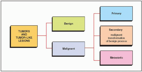
FIGURE 16.1 Classification of tumors and tumor-like lesions.


TABLE 16.1 Classification of Tumors and Tumor-like Lesions by Tissue of Origin


Tissue of Origin


Benign Lesion


Malignant Lesion


Bone forming (osteogenic)


Osteoma


Osteosarcoma (and variants)


Osteoid osteoma


Juxtacortical osteosarcoma (and variants)


Osteoblastoma


Cartilage forming (chondrogenic)


Enchondroma (chondroma)


Chondrosarcoma (central)


Periosteal (juxtacortical) chondroma


Conventional


Enchondromatosis (Ollier disease)


Mesenchymal


Osteochondroma (osteocartilaginous exostosis, solitary or multiple)


Clear cell


Dedifferentiated


Chondroblastoma


Chondrosarcoma (peripheral)


Chondromyxoid fibroma


Periosteal (juxtacortical)


Fibrocartilaginous mesenchymoma


Fibrous, osteofibrous, and fibro-histiocytic (fibrogenic)


Fibrous cortical defect (metaphyseal fibrous defect)


Fibrosarcoma


Nonossifying fibroma


Malignant fibrous histiocytoma


Benign fibrous histiocytoma


Fibrous dysplasia (monostotic and polyostotic)


Fibrocartilaginous dysplasia


Focal fibrocartilaginous dysplasia of long bones


Periosteal desmoid


Desmoplastic fibroma


Osteofibrous dysplasia (Kempson-Campanacci lesion)


Ossifying fibroma (Sissons lesion)


Vascular


Hemangioma


Angiosarcoma


Glomus tumor


Hemangioendothelioma


Cystic angiomatosis


Hemangiopericytoma


Hematopoietic, reticuloendothelial, and lymphatic


Giant cell tumor (osteoclastoma)


Malignant giant cell tumor


Langerhans cell histiocytosis


Histiocytic lymphoma


Lymphangioma


Hodgkin lymphoma


Leukemia


Myeloma (plasmacytoma)


Ewing sarcoma


Neural (neurogenic)


Neurofibroma


Malignant schwannoma


Neurilemoma


Neuroblastoma


Primitive neuroectodermal tumor (PNET)


Chordoma


Notochordal


Lipoma


Liposarcoma


Fat (lipogenic)


Simple bone cyst


Adamantinoma


Unknown


Aneurysmal bone cyst


Intraosseous ganglion


TABLE 16.2 Benign Conditions With Potential for Malignant Transformation


Benign Lesion


Malignancy


Enchondroma (in the long or flat bones*; in the short, tubular bones almost always as a part of Ollier disease or Maffucci syndrome)


Chondrosarcoma


Osteochondroma


Peripheral chondrosarcoma


Synovial chondromatosis


Chondrosarcoma


Fibrous dysplasia (usually polyostotic, or treated with radiation)


Fibrosarcoma


Malignant fibrous histiocytoma


Osteosarcoma


Osteofibrous dysplasia (Kempson-Campanacci lesion)


Adamantinoma


Neurofibroma (in plexiform neurofibromatosis) Malignant schwannoma


Liposarcoma


Malignant mesenchymoma


Medullary bone infarct


Fibrosarcoma


Malignant fibrous histiocytoma


Osteomyelitis with chronic draining sinus tract (usually more than 15-20 years duration)


Squamous cell carcinoma


Fibrosarcoma


Paget disease


Osteosarcoma


Chondrosarcoma


Fibrosarcoma


Malignant fibrous histiocytoma


* Some authorities believe that, at least in some “malignant transformations” of enchondroma to chondrosarcoma, there was in fact from the very beginning a malignant lesion masquerading as benign and not recognized as such.

Some authorities believe that this is not a true malignant transformation, but rather independent development of malignancy in the benign condition.


Computed Tomography

Although CT by itself is rarely helpful in making a specific diagnosis, it can provide a precise evaluation of the extent of a bone lesion and may demonstrate breakthrough of the cortex and involvement of surrounding soft tissues (Fig. 16.7). CT is moreover very helpful in delineating a bone tumor having a complex anatomic structure. The scapula (Fig. 16.8), pelvis (Fig. 16.9), and sacrum, for example, may be difficult to image fully with conventional radiographic techniques. At times, three-dimensional computed tomography (3D CT)-reconstructed images are used to better and more comprehensively demonstrate the tumors. This technique can be useful, for example, in depicting surface lesions of bone, such as osteochondroma (Fig. 16.10), parosteal osteosarcoma, or juxtacortical chondrosarcoma. CT examination is crucial in determining the extent and spread of a tumor in the bone if limb salvage is contemplated, so that a safe margin of resection can be planned (Fig. 16.11). It can effectively demonstrate the intraosseous extension of a tumor and its extraosseous involvement of soft tissues such as muscles and neurovascular bundles. CT is also useful for monitoring the results of treatment, evaluating for recurrence of a resected tumor, and demonstrating the effect of nonsurgical treatment such as radiation therapy or chemotherapy (Fig. 16.12). It is also helpful in evaluating soft-tissue tumors (Fig. 16.13), which on standard radiographs are indistinguishable from one another (with the exception of lipomas, which usually demonstrate low-density features), blending imperceptibly into the surrounding normal tissue.


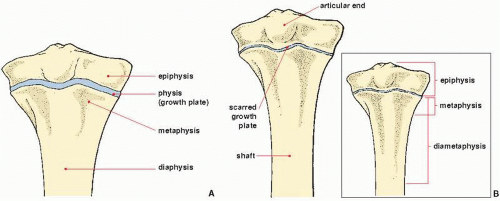
FIGURE 16.2 Parts of the bone. (A) In the maturing skeleton, the epiphysis, growth plate, metaphysis, and diaphysis are clearly recognizable areas. (B) With skeletal maturity, distinct epiphyseal and metaphyseal zones have ceased to exist. The terminology for describing the location of lesions should alter accordingly. The inset illustrates an alternate terminology.


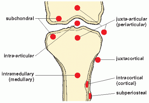
FIGURE 16.3 Terminology used to describe the location of lesions in the bone.

Contrast enhancement of CT images aids in the identification of major neurovascular structures and well-vascularized lesions. Evaluating the relationship between the tumor and the surrounding soft tissues
and neurovascular structures is particularly important for planning limb-salvage surgery.


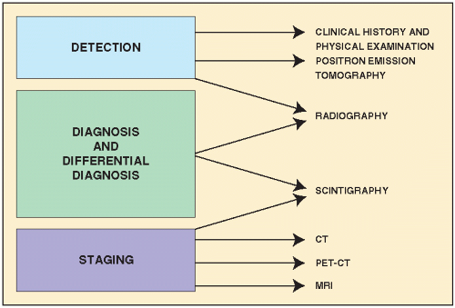
FIGURE 16.4 Imaging of tumors. Imaging of musculoskeletal neoplasms can be considered from three aspects: detection, diagnosis and differential diagnosis, and staging. (Modified from Greenspan A et al., 2007.)


PET and PET-CT

Recently, 2-fluoro[fluorine-18]-2-deoxy-D-glucose (F-18 FDG) PET and PET-CT have emerged as very effective metabolic-anatomic imaging techniques for the assessment of variety of neoplastic conditions. The simultaneous detection and precise localization of metabolic and biochemical activities by PET combined with anatomic details obtained by CT into a single superimposed image provides the radiologist with an unique opportunity not only to make a distinction between the normal and pathologic processes, but frequently between the various pathologic disorders as well. Although the most common use of PET/CT is to improve the staging of musculoskeletal tumors and evaluate their response to therapy and emergence of recurrences, this technique is also a powerful tool for the detection and evaluation of metastatic disease (Fig. 16.14, see also Figs. 2.28B and 2.31) and some primary musculoskeletal tumors (Fig. 16.15, see also Figs. 2.29 and 2.30). In addition, recent trials using dual-time point F-18 FDG PET to distinguish malignant tumors from benign conditions yielded promising results.


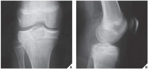
FIGURE 16.5 Chondroblastoma. Anteroposterior (A) and lateral (B) radiographs of the right knee of a 13-year-old girl reveal a radiolucent lesion located eccentrically in the proximal epiphysis of the tibia, with sharply defined borders and a thin, sclerotic margin. Here, the standard projections led to the correct radiographic diagnosis of chondroblastoma.


Arteriography

Arteriography is used mainly to map out bone lesions and to assess the extent of disease. It is also used to demonstrate the vascular supply of a tumor and to locate vessels suitable for preoperative intra-arterial chemotherapy, as well as to demonstrate the area suitable for open biopsy because the most vascular area of a tumor contains the most aggressive
component. Occasionally, arteriography can be used to demonstrate abnormal tumor vessels, corroborating findings with conventional radiography (Fig. 16.16). Arteriography is often useful in planning for limb-salvage procedures because it demonstrates the regional vascular anatomy and thus permits a plan to be drawn up for the resection procedure. It is also sometimes used to outline the major vessels before resection of a benign tumor (Fig. 16.17), and it can be combined with an interventional procedure, such as embolization of hypervascular tumors, before further treatment (Fig. 16.18). In selected cases, arteriography may help make a differential diagnosis, such as of osteoid osteoma versus a bone abscess.


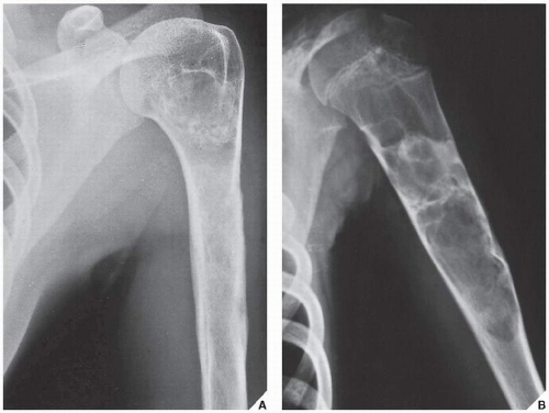
FIGURE 16.6 Comparison radiography: a simple bone cyst. (A) Anteroposterior radiograph of the left humerus in a 26-year-old woman with vague pain for 2 months shows an ill-defined lesion in the medullary region, with a periosteal reaction medially and laterally. There appear to be scattered calcifications in the proximal portion of the lesion. The possibility of a cartilage tumor such as chondrosarcoma was considered, but a radiograph taken 17 years earlier (B) shows an unquestionably benign lesion (a simple bone cyst) that had been treated by curettage and the application of bone chips. In view of this, the later findings were interpreted as representing a healed bone cyst. The patient’s pain was found to be related to muscle strain.


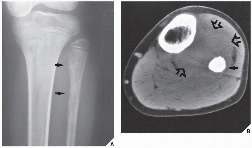
FIGURE 16.7 Ewing sarcoma. (A) Anteroposterior radiograph demonstrates a malignant lesion that proved to be Ewing sarcoma in the proximal diaphysis of the left fibula (arrows) of a 12-year-old boy. (B) On CT examination, there is involvement of the bone marrow (arrow) and extension of the tumor into the soft tissues (open arrows).


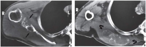
FIGURE 16.8 Chondrosarcoma. Standard radiographs were ambiguous in this 70-year-old man with a palpable mass over the right scapula. However, two CT sections demonstrate a destructive lesion of the glenoid portion and body of the scapula (arrows) (A), with a large soft-tissue mass extending to the rib cage and containing calcifications (curved arrows) (B).


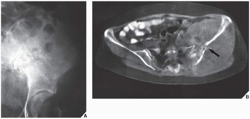
FIGURE 16.9 Osteosarcoma. (A) Standard anteroposterior radiograph of the pelvis was not sufficient to delineate the full extent of the destructive lesion of the iliac bone in this 66-year-old woman. (B) A CT scan, however, showed a pathologic fracture of the ilium (arrow) and the full extent of soft-tissue involvement. The high Hounsfield values of the multiple soft-tissue densities suggested bone formation. Enhancement of the CT images with contrast agent showed an increased vascularity of the lesion. Collectively, the CT findings suggested a diagnosis of osteosarcoma that, although unusual for a person of this age, was confirmed by open biopsy.


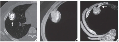
FIGURE 16.10 Osteochondroma: effectiveness of 3D CT. (A) Conventional CT section through the chest shows an osteochondroma at the site of the anterolateral portion of the right forth rib (arrow). It is difficult to determine if the lesion is sessile or pedunculated. (B) 3D CT reconstructed image in maximum intensity projection (MIP) delivers a much more informative image of osteochondroma, and allows one to characterize the internal architecture of the lesion; note typical chondroid matrix of the tumor. (C) 3D CT reconstructed image in shaded surface display (SSD) renders better conspicuity of the lesion; the pedicle of osteochondroma (arrow) is now clearly demonstrated. (Reprinted from Greenspan A et al., 2007.)


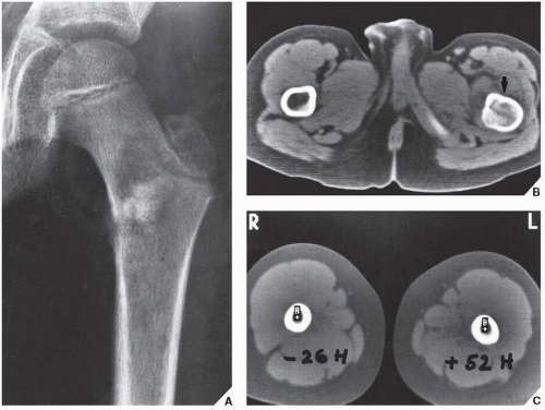
FIGURE 16.11 Osteosarcoma: effectiveness of CT. (A) Anteroposterior radiograph of the left proximal femur of a 12-year-old boy demonstrates an osteolytic lesion in the intertrochanteric region, with a poorly defined margin and amorphous densities in the center associated with a periosteal reaction medially—features suggesting osteosarcoma, which was confirmed on open biopsy. Because a limb-salvage procedure was contemplated, a CT scan was performed to determine the extent of marrow infiltration and the required level of bone resection. The most proximal section (B) shows obvious gross tumor involvement of the marrow cavity of the left femur (arrow). A more distal section (C) shows no gross marrow abnormality, but a positive Hounsfield value of 52 units indicates tumor involvement of the marrow, which was not shown on the standard radiographs. By comparison, the section of the right femur shows a normal Hounsfield value of -26 for bone marrow.


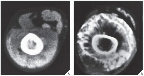
FIGURE 16.12 Osteosarcoma after chemotherapy. Before surgery, this 14-year-old girl with an osteosarcoma of the left femur underwent a full course of chemotherapy. (A) CT section before the therapy was begun shows involvement of the bone and marrow cavity. Note the soft-tissue extension of the tumor, with nonhomogeneous, amorphous tumor bone formation. After combined treatment with doxorubicin hydrochloride, vincristine, methotrexate, and cisplatin, a repeat CT scan (B) shows calcifications and ossifications in the periphery of the lesion, which represents reactive rather than tumor bone and demonstrates the success of chemotherapy. Radical excision of the femur and a subsequent histopathologic examination showed almost complete eradication of malignant cells, confirming the CT findings.


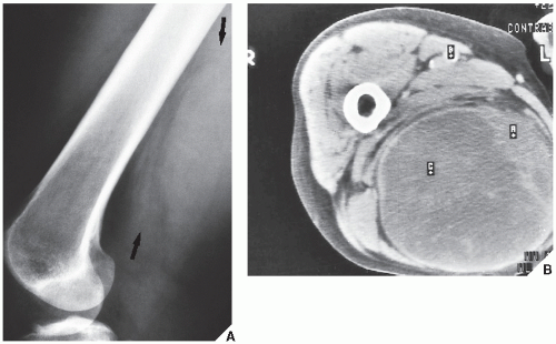
FIGURE 16.13 CT of malignant fibrous histiocytoma (MFH) of the soft tissue. A 56-year-old woman presented with a soft-tissue mass on the posteromedial aspect of the right thigh. (A) Lateral radiograph of the femur demonstrates only a soft-tissue prominence posteriorly (arrows). (B) CT section shows an axial image of the mass, which is contained by a fibrotic capsule. The overlying skin is not infiltrated. Despite the benign appearance, the mass proved on biopsy to be a MFH.


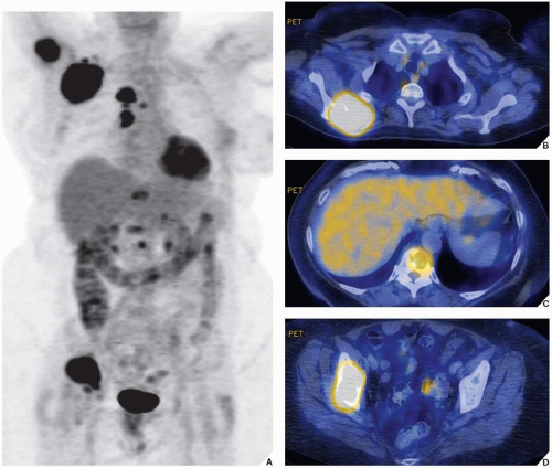
FIGURE 16.14 PET and PET-CT of metastases. A 61-year-old woman was diagnosed with lung carcinoma. (A) A whole body PET scan shows several hypermetabolic foci in the internal organs, lymph nodes, and osseous structures, representing metastatic disease. The fused PET-CT images demonstrate metastatic lesions in the right scapula (B), thoracic vertebral body (C), and right ilium (D).


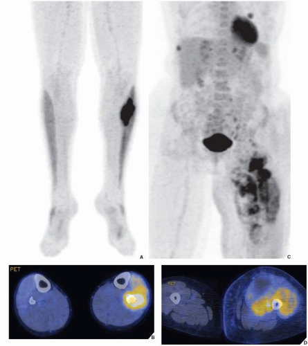
FIGURE 16.15 PET and PET-CT of primary bone and primary soft-tissue tumors. (A,B) A hypermetabolic focus in the proximal left fibula in a 23-year-old man proved to be a Ewing sarcoma. (C,D) A hypermetabolic lesion in the vastus lateralis and medialis in the proximal left thigh in a 58-year-old woman was diagnosed on histopathologic examination as MFH of the soft tissues.


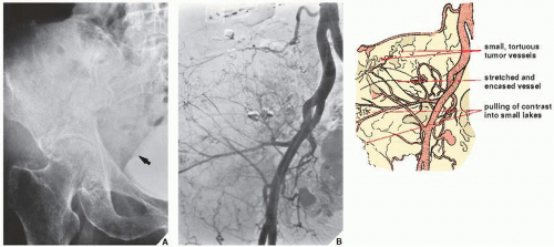
FIGURE 16.16 Arteriography of dedifferentiated chondrosarcoma. (A) Anteroposterior radiograph of the pelvis in a 79-year-old woman with an 8-month history of pain in the right buttock and weight loss demonstrates a poorly defined destructive lesion of the right iliac bone, with multiple small calcifications and a soft-tissue mass extending into the pelvis. Note the effect of the mass on the urinary bladder filled with contrast (arrow). A chondrosarcoma was suspected, and a femoral arteriogram was performed as part of the diagnostic workup. (B) Subtraction study of an arteriogram demonstrates hypervascularity of the tumor. Note the abnormal tumor vessels, encasement and stretching of some vessels, and “pulling” of contrast medium into small “lakes”—all characteristic signs of a malignant lesion. Biopsy revealed a highly malignant, dedifferentiated chondrosarcoma. In this case, the vascular study corroborated the radiographic findings of a malignant bone tumor.


Myelography

Myelography may be helpful in dealing with tumors that invade the vertebral column and thecal sac (Fig. 16.19), although recently this procedure has been almost completely replaced by MRI.


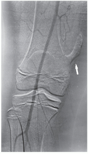
FIGURE 16.17 Arteriography of osteochondroma. A 12-year-old boy with osteochondroma of the distal femur (arrow) underwent arteriography to demonstrate the relationship of the distal superficial femoral artery to the lesion. This subtraction study shows no major vessels near the planned site of resection at the base of the lesion, important information for surgical planning.


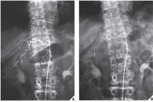
FIGURE 16.18 Vertebral arteriography and embolization of hemangioma. A 73-year-old woman presented with a collapsed T11 vertebra, which showed a corduroy-like pattern suggestive of hemangioma. Vertebral angiography was performed. (A) Arteriogram of the 11th right intercostal artery outlines a vascular paraspinal mass associated with hemangioma and indicating extension of the lesion into the soft tissues. (B) After embolization, the lesion shows a marked decrease in vascularity. Subsequently, the patient underwent decompression laminectomy and anterior fusion at T10-11 using a fibular strut graft.


Magnetic Resonance Imaging

MRI is indispensable in evaluating bone and soft-tissue tumors. Particularly with soft-tissue masses, MRI offers distinct advantages over CT. There is improved visualization of tissue planes surrounding the lesion, for example, and neurovascular involvement can be evaluated without the use of intravenous contrast.

In the evaluation of intraosseous and extraosseous extensions of a tumor, MRI is crucial because it can determine with high accuracy the presence or absence of soft-tissue invasion by a tumor (Fig. 16.20). MRI has often proved to be superior to CT in delineating the extraosseous and intramedullary extent of the tumor and its relationship to surrounding structures (Fig. 16.21). By showing sharper demarcation between normal and abnormal tissue than CT, MRI—particularly in evaluation of the extremities—reliably identifies the spatial boundaries of tumor masses (Fig. 16.22), the encasement and displacement of major neurovascular bundles, and the extent of joint involvement. Spin-echo
T1-weighted images enhance tumor contrast with bone, bone marrow, and fatty tissue, whereas spin-echo T2-weighted images enhance tumor contrast with muscle and accentuate peritumoral edema. Axial and coronal images have been used in determining the extent of soft-tissue invasion in relation to important vascular structures. However, in comparison with CT, MR images do not clearly demonstrate calcification in the tumor matrix; in fact, large amounts of calcification or ossification may be almost undetectable. Moreover, MRI has been shown to be less satisfactory than CT in the demonstration of cortical destruction. It is important to realize that both MRI and CT have advantages and disadvantages, and circumstances exist in which either can be the preferential or complementary study. But it is even more important that the surgeon
tell the radiologist who is performing and interpreting the study what information is needed.


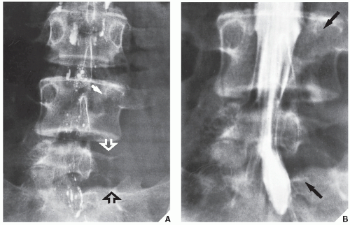
FIGURE 16.19 Myelography of aneurysmal bone cyst. Initial radiographic examination of the lumbar spine of this 14-year-old girl with an 18-month history of pain in the lower back and sciatica of the left leg did not disclose any abnormalities; myelography was performed because of suspected herniation of a lumbar disk, but it was inconclusive. A repeat study was requested when the symptoms became more severe after 3 months. (A) Posteroanterior radiograph of the lumbosacral spine shows destruction of the left pedicle of L-4 (arrow) and the left part of the L5 body (open arrows). Note the residual contrast in the subarachnoid space. A repeat myelogram using a water-soluble contrast (metrizamide) shows, on the posteroanterior view (B), extradural compression of the thecal sac on the left side with displacement of the nerve roots (arrows). Biopsy confirmed the radiographic diagnosis of an aneurysmal bone cyst.


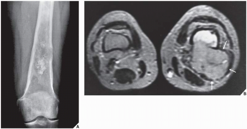
FIGURE 16.20 MRI of chondrosarcoma. (A) Conventional radiograph of the left femur in anteroposterior projection of a 67-year-old woman with chondrosarcoma demonstrates a tumor in the distal shaft destroying the medullary portion of the bone and breaking through the cortex. The soft-tissue extension cannot be determined. (B) Axial T2-weighted MR image (SE; TR 2500/TE 70 msec) demonstrates a tumor infiltrating bone marrow, destroying the posterolateral cortex, and breaking into the soft tissues with the formation of a large mass (arrows). Compare with a normal contralateral extremity.


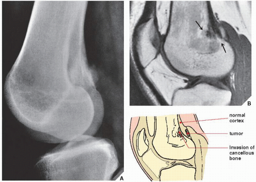
FIGURE 16.21 MRI of parosteal osteosarcoma. (A) From this lateral radiograph of the distal femur of a 22-year-old woman with parosteal osteosarcoma, it is difficult to evaluate if the tumor is on the surface of the bone or already infiltrated through the cortex. (B) Sagittal T1-weighted MRI (SE; TR 500/TE 20 msec) demonstrates invasion of the cancellous portion of the bone, as represented by an area of low signal intensity (arrows).


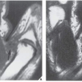
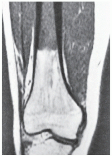
FIGURE 16.22 MRI of MFH. Coronal T1-weighted MRI (SE; TR 500/TE 20 msec) demonstrates involvement of the medullary cavity of the right femur in this 16-year-old girl with MFH. Note the excellent demonstration of the interface between normal bone displaying high-signal intensity and a tumor displaying intermediate signal intensity.

Several investigators have stressed the superior contrast enhancement of MR images using intravenous injection of gadopentetate dimeglumine (gadolinium diethylenetriamine-penta-acetic acid, [Gd-DTPA]). Enhancement was found to give better delineation of the tumor’s richly vascularized parts and of the compressed tissue immediately surrounding the tumor. It was also found to assist in the differentiation of intraarticular tumor extension from joint effusion, and, as Erlemann pointed out, improved the differentiation of necrotic tissue from viable areas in various malignant tumors.

According to the recent investigations, MRI may have an additional application in evaluating both the tumor’s response to radiation and chemotherapy and any local recurrence. On gadolinium-enhanced T1-weighted images, signal intensity remains low in avascular, necrotic areas of tumor while it increases in viable tissue. Although static MRI was of little value for the assessment of response to the treatment, dynamic MRI using Gd-DTPA as a contrast enhancement, according to Erlemann, had the highest degree of accuracy (85.7%) and was superior to scintigraphy, particularly in patients who were receiving intra-arterial chemotherapy. In general, drug-sensitive tumors display slower uptake of Gd-DTPA after preoperative chemotherapy than do nonresponsive lesions. As Vaupel contended, the rapid uptake of Gd-DTPA by malignant tissues may be due to increased vascularity and more rapid perfusion of the contrast material through an expanded interstitial space. The latest observation by Dewhirst and Kautcher suggests that MR spectroscopy may also be useful in the evaluation of patients undergoing chemotherapy.

It must be stressed, however, that most of the time MRI is not suitable for establishing the precise nature of a bone tumor. In particular, too much faith has been placed in MRI as a method of distinguishing benign lesions from malignant ones. An overlap between the classic characteristics of benign and malignant tumors is often observed. Moreover, some malignant bone tumors can appear misleadingly benign on MR images and, conversely, some benign lesions may exhibit a misleadingly malignant appearance. Attempts to formulate precise criteria for correlating MRI findings with histologic diagnosis have been largely unsuccessful. Tissue characterization on the basis of MRI signal intensities is still unreliable. Because of the wide spectrum of bone tumor composition and their differing histologic patterns, as well as in tumors of similar histologic diagnosis, signal intensities of histologically different tumors may overlap or there may be variability of signal intensity in histologically similar tumors.

Trials using combined hydrogen-1 MRI and P-31 MR spectroscopy also failed to distinguish most benign lesions from malignant tumors. Despite the use of various criteria, the application of MRI to tissue diagnosis has rarely brought satisfactory results. This is because, in general, the small number of protons in calcified structures renders MRI less effective in diagnosing bone lesions, and hence valuable evidence concerning the production of the tumor matrix can be missed. Moreover, as several investigations have shown, MRI is an imaging modality of low specificity. T1 and T2 measurements are generally of limited value for histologic characterization of musculoskeletal tumors. Quantitative determination of relaxation times has not proved to be clinically valuable in identifying various tumor types, although, as noted by Sundaram, it has proved to be an important technique in the staging of osteosarcoma and chondrosarcoma. T2-weighted images in particular are a crucial factor in delineating extraosseous tumor extension and peritumoral edema, as well as in assessing the involvement of major neurovascular bundles. Necrotic areas change from a low-intensity signal in the T1-weighted image to a very bright, intense signal in the T2-weighted image and can be differentiated from viable, solid tumor tissue. Although MRI cannot predict the histology of bone tumors, as Sundaram pointed out, it is a useful tool for distinguishing round cell tumors and metastases from stress fractures or medullary infarcts in symptomatic patients with normal radiographs, and, according to Baker, it can occasionally differentiate benign from pathologic fracture.


Skeletal Scintigraphy

The radionuclide bone scan is an indicator of mineral turnover, and because there is usually enhanced deposition of bone-seeking radiopharmaceuticals in areas of bone undergoing change and repair, a bone scan is useful in localizing tumors and tumor-like lesions in the skeleton, particularly in such conditions as fibrous dysplasia, Langerhans cell histiocytosis, or metastatic cancer, in which more than one lesion is encountered (Fig. 16.23). It also plays an important role in localizing small lesions such as osteoid osteomas, which may not always be seen on conventional radiographs (see Fig. 17.11B). Although in most instances, a radionuclide bone scan cannot distinguish benign lesions from malignant tumors, because increased blood flow with increased isotope deposition and increased osteoblastic activity takes place in benign and malignant conditions, it is still occasionally capable of making such differentiation in benign lesions that do not absorb the radioactive isotope (Fig. 16.24). The radionuclide bone scan is sometimes also useful for differentiating multiple myeloma, which usually shows no significant uptake of the tracer, from metastatic cancer, which usually does.

Aside from routine radionuclide scans performed using 99mTc-labeled phosphate compounds, occasionally 67Ga is used for the detection and staging of bone and soft-tissue neoplasms. Gallium is handled by the body much like iron in that the protein transferrin carries it in the plasma, and it also competes for extravascular iron-binding proteins such as lactoferrin. The administered dose for adults ranges from 3 mCi (111 MBq) to 10 mCi (370 MBq) per study. The exact mechanism of tumor uptake of gallium remains unsettled, and its uptake varies with tumor type. In particular, Hodgkin lymphomas and histiocytic lymphomas are prone to significant gallium uptake.


Interventional Procedures

Percutaneous bone and soft-tissue biopsy performed in the radiology department has in recent years gained its place in the diagnostic workup for various neoplastic diseases, including bone tumors. In patients with primary bone neoplasms, it is a helpful diagnostic and evaluative tool,
allowing rapid histologic diagnosis, which is now considered essential, particularly in the planning of a limb-salvage procedure. It also helps assess the effect of chemotherapy and radiation therapy and helps locate the site of the primary tumor in cases of metastatic disease (Fig. 16.25). In addition, percutaneous bone and soft-tissue biopsy performed in the radiology suite is simpler and costs less than a biopsy performed in the operating room.


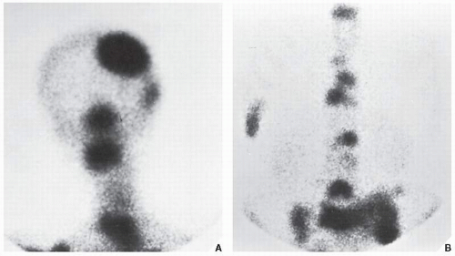
FIGURE 16.23 Scintigraphy of the metastases. A radionuclide bone scan was performed on a 68-year-old woman with metastatic breast carcinoma to determine the distribution of metastases. After an intravenous injection of 15 mCi (555 MBq) of 99mTc diphosphonate, an increased uptake of the radiopharmaceutical agent is seen in the skull and cervical spine (A) and lumbar spine and pelvis (B), localizing the site of the multiple metastases.


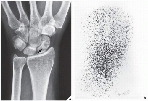
FIGURE 16.24 Scintigraphy of enostosis. A 32-year-old woman presented with pain localized in the wrist area. (A) Dorsovolar radiograph of the wrist demonstrates a sclerotic round lesion in the scaphoid (arrow), and a diagnosis of osteoid osteoma was considered. (B) Radionuclide bone scan reveals normal isotope uptake, ruling out osteoid osteoma, which is invariably associated with an increased uptake of radiopharmaceutical. The lesion instead proved to be a bone island (enostosis), an asymptomatic developmental error of endochondral ossification without any consequence to the patient. The pain was unrelated to the island, coming instead from tenosynovitis; it disappeared after the patient was treated for the latter condition.


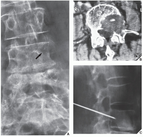
FIGURE 16.25 Percutaneous bone biopsy. (A) Anteroposterior radiograph of the lumbar spine in a 67-year-old woman with lower back pain for 4 months demonstrates destruction of the left pedicle of the L4 vertebra (arrow). (B) CT section shows, in addition, involvement of the vertebral body by the tumor. (C) Percutaneous biopsy of the lesion, performed in the radiology suite for the purpose of rapid histopathologic diagnosis, revealed a metastatic adenocarcinoma from the colon.


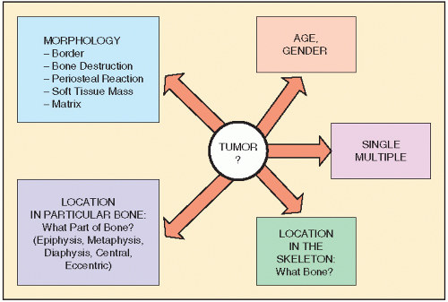
FIGURE 16.26 Diagnosis of bone lesion. Analytic approach to evaluation of the bone neoplasm must include patient age, multiplicity of a lesion, location in the skeleton and in the particular bone, and radiographic morphology.


Tumors and Tumor-like Lesions of the Bone


Diagnosis

Patient age and determination of whether a lesion is solitary or multiple are the starting approaches in the diagnosis of bone tumors (Fig. 16.26).


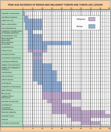
FIGURE 16.27 Peak age incidence of benign and malignant tumors and tumor-like lesions. (Sources: Dahlin DC, 1986; Dorfman HD, Czerniak B, 1998; Fechner RE, Mills SE, 1993; Huvos AG, 1979; Jaffe HL, 1968; Mirra JM, 1989; Moser RP, 1990; Schajowicz F, 1994; Unni KK, 1988; Wilner D, 1982.)


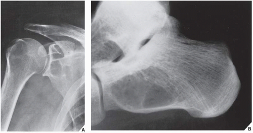
FIGURE 16.28 Simple bone cyst. (A) Anteroposterior radiograph of the right shoulder of a 69-year-old man with shoulder pain for 8 months demonstrates a well-defined radiolucent lesion with a sclerotic border in the glenoid portion of the scapula. Because the patient had a history of gout, the lesion was thought to represent an intraosseous tophus. In the differential diagnosis, an intraosseous ganglion and even a cartilage tumor were also considered. An excision biopsy, however, revealed a simple bone cyst, which is very unusual in the glenoid part of the scapula. (B) Lateral radiograph of the left hindfoot of a 50-year-old woman shows a radiolucent lesion in the calcaneus proven on the excision biopsy to be a simple bone cyst.


Clinical Information

The age of the patient is probably the single most important item of clinical data in radiographically establishing the diagnosis of a tumor (Fig. 16.27). Certain tumors have a predilection for specific age groups. Aneurysmal bone cysts, for example, rarely occur beyond age 20, and giant cell tumors as a rule are found only after the growth plate is closed. Other lesions may have different radiographic presentations or occur in different locations in patients of different ages. Simple bone cysts, which before skeletal maturity present almost exclusively in the long bones such as the proximal humerus and proximal femur, may appear in other locations (pelvis, scapula, os calcis) and have unconventional radiographic presentations with progressing age (Fig. 16.28).

Also important for clinically differentiating lesions of similar radiographic presentation—such as Langerhans cell histiocytosis (formerly called eosinophilic granuloma), osteomyelitis, and Ewing sarcoma—is the duration of the patient’s symptoms. In Langerhans cell histiocytosis, for example, the amount of bone destruction seen radiographically after 1 week of symptoms is usually the same as that seen after 4 to 6 weeks of symptoms in osteomyelitis and 3 to 4 months in Ewing sarcoma.

Occasionally, race may also be an important differential diagnostic factor because certain lesions, such as tumoral calcinosis or bone infarctions, are seen more commonly in blacks than in whites, whereas others, such as Ewing sarcoma, are almost never seen in blacks.

The growth rate of the tumor may be an additional factor in differentiating malignant tumors (usually rapid-growing) from benign tumors (usually slow-growing).

Laboratory data, such as an increased erythrocyte sedimentation rate or an elevated alkaline or acid phosphatase level in the serum, occasionally can be a corroborative factor in diagnosis.


Imaging Modalities

With so many imaging techniques available to diagnose and characterize the bone tumor further, radiologists and clinicians are frequently at a loss as to how to proceed in a given case, what modality to use for this particular problem, in what order of preference to use the modalities, and when to stop. It is important to keep in mind that the choice of techniques for imaging the bone or soft-tissue tumor should be dictated not only by the clinical presentation and the technique’s expected effectiveness but also by equipment availability, expertise, cost, and restrictions applicable to individual patients (e.g., allergy to ionic or nonionic iodinated contrast agents may preclude the use of arthrography; presence of a pacemaker may preclude the use of MRI; or physiologic states such as pregnancy warrant the use of ultrasound over the use of ionized radiation). Some of these problems were discussed in general in Chapters 1 and 2.

Only gold members can continue reading. Log In or Register to continue

Stay updated, free articles. Join our Telegram channel

Jul 24, 2016 | Posted by in MUSCULOSKELETAL IMAGING | Comments Off on Radiologic Evaluation of Tumors and Tumor-like Lesions

Full access? Get Clinical Tree

Get Clinical Tree app for offline access