Fig. 17.1
RTRT system in Hokkaido University. Black arrow shows the image intensifier (I.I.)
We have reported the clinical results of stereotactic body radiation therapy (SBRT) for non-small cell lung cancer (NSCLC), hepatocellular carcinoma (HCC), adrenal tumor, and spinal schwannoma [5–8]. Analysis of marker motion was also done and several interesting findings were reported [9–12].
In this article, we describe experiences about gold marker insertion, clinical results of SBRT using RTRT system, analysis of marker movements, and the future direction of RTRT system.
17.2 The Method of Gold Marker Insertion
17.2.1 Non-small Cell Lung Cancer
The method of implantation of gold marker into lung is described by Harada et al. [13] and Imura et al. [14]. They used bronchoscope and catheter to insert gold markers. Gold marker was inserted into catheter and pushed by hard plastic wire. The gold marker was inserted and wedged into the bronchial trees near the tumor. The location of bronchoscope, catheter, and markers were visualized by fluoroscopy [13].
Imura et al. reported that 94 % of markers that were seen in CT for radiotherapy planning were detected throughout the radiotherapy delivery [14]. They also reported that the 95 % of distance variation between implanted markers was within 2 mm. The location of tumor and marker fixation rate was correlated. The fixation rate in the left upper lobe was lower than in the other lobes. They suggested that learning curve existed. Pneumothorax was found in only 1 of 57 patients [14].
Imura et al. reported the histological change after marker insertion. They found that fibrotic change was seen after 5–7 days after marker insertion [15]. They suggested that radiotherapy should be started above 5 days after gold marker insertion because marker dislocation tended not to occur due to fibrotic change around the marker.
17.2.2 Hepatocellular Carcinoma
Gold marker was also able to insert into liver and prostate [16, 17]. Implantation of the marker into liver through skin was done under ultrasound and/or CT guidance. The implantation kit for spinal lesions and prostate and liver tumors was manufactured by Medikit Co. Ltd. (Tokyo, Japan). There was only 1 migration of 21 implanted markers in liver. Shirato et al. reported that no serious complication due to marker implantation was observed [16]. However, we experienced transient bile ductal bleeding in 1 patient in SBRT for HCC after the report of Taguchi et al. [6].
17.3 CT Acquisition and Treatment Planning
CT for treatment planning was performed 3 days after insertion of gold marker in general. Patients were asked to hold breath at exhale phase during CT acquisition. Slice thickness was 2.5 mm in principle.
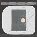
Gross tumor volume and clinical target volume (CTV) was made as other SBRT methods without RTRT system. Markers were also contoured. Gating window was usually 2 mm, but this was changed to 3 mm by respiratory irregularity in actual treatment. PTV was made by adding margin of gating window and other uncertainty to CTV, the margin was usually 5 mm. Six beams, 4 coplanar and 2 non-coplanar beams, were typically selected for treatment in Hokkaido University Hospital. Figure 17.2 shows the flow chart from the assessment of indication to the treatment.
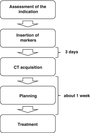
Fig. 17.2
Flow chart of treatment with RTRT system
17.4 Clinical Results
17.4.1 Non-small Cell Lung Cancer
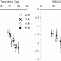
Inoue et al. reported the results of stereotactic body radiation therapy (SBRT) using RTRT system for non-small cell lung cancer (NSCLC) [5]. They used superposition algorithm for inhomogeneity correction and 48 Gy in 4 fractions at isocenter or 40 Gy in 4 fractions at D95 of PTV. The 5-year local control rate (LC) was 78 % (95 % confidence interval (CI), 68–90 %) and the 5-year overall survival rate (OS) was 64 % (95 % CI, 53–78 %), respectively. The 5 year overall survival (OS) in patients with T1a was 75 %. The 5 year OS in patients with T1b or T2 (p = 0.01) was 56 %, which was statistically worse than that of patients with T1a (P = 0.01). They also reported that the maximum amplitude of marker movement in lower lobe was significantly larger than that in upper lobe; however, there was no significant difference in LC between the lower lobe and the other lobe. The fact that no difference existed in LC between the lower and the other lobes shows that we treat properly the moving tumor using RTRT system.
17.4.2 Hepatocellular Carcinoma
Taguchi et al. reported the results of SBRT using RTRT system for 18 lesions of HCC [6]. Various dose fractionation schedule was used, and 48 Gy in 8 fractions at isocenter was mostly used (n = 7). A symptomatic complication due to the insertion of the fiducial marker occurred in 1 patient, who experienced transient bile ductal bleeding and inflammation. The 2 year OS was 44 % after SBRT and the LC within the CTV was 83 % at 30 months after SBRT. The intrahepatic control rate was 17 % at 2 years after SBRT. An adverse reaction due to radiation was seen in 2 patients. A transient gastric ulcer was occurred in 1 patient and radiation pneumonitis was occurred in 1 patient, respectively.
This result is difficult to interpret. Patients were highly selected, HCC shows multicentric nature, and dose was varied. Adding to that, several local treatments are effective for HCC. Surgery, RFA, and TACE is major therapy in Japan. Radiotherapy is not recognized as standard first treatment option for HCC. However, radiotherapy like SBRT or proton beam therapy is reported effective in retrospective studies; SBRT may be regarded as one of the effective local treatment based on future clinical trials.
17.4.3 Other Tumors
Katoh et al. reported the results of the treatment for metastatic adrenal tumor using RTRT system [7]. They treated 10 lesions with 48 Gy in 8 fractions and 1 lesion with 30 Gy in 8 fractions. They reported that no symptomatic adverse effects were observed and the actuarial freedom-from local progression rate was 100 % at 12 months. The concept of oligometastases changed the treatment policy for patients with small number of metastatic lesions [18]. Curative dose for metastatic adrenal tumor may be beneficial for the selected patients with metastatic adrenal tumor.
Onimaru et al. reported that the experience the radiotherapy treatment for spinal schwannoma using RTRT system [8]. They calculated the rotation using 3 markers. It seems to be important to consider the rotation and the deformation of target, however, the strategy to the rotation and the deformation is still to be investigated.
17.5 Marker Movement Analysis
RTRT system records log file about marker motion. The log file is very valuable because it is the exact data during irradiation delivery. Several investigators reported the results of analyzing log files of RTRT system and showed interesting results [9–12].
Seppenwoolde et al. developed analyzing system and reported the marker motion in the lung [9]. This report showed that respiratory movement of marker shows a hysteresis. It was the first report about baseline shift during the radiation delivery and intra-fractional error based on data during irradiation delivery [9].
Onimaru et al. reported the relationship between the marker location and the amplitude of marker movement in NSCLC [10]. They showed that marker movement for the craniocaudal direction was smaller in the anterior part and the cranial part of lung.
Onodera et al. evaluated the relationship between lung parenchymal findings on CT and the amplitude of marker movement [11]. They found that presence of fibrosis and pleural tumor contacts were weakly associated with marker motion. There was no correlation between lung fibrosis and marker motion in the lower lung. There was also no correlation between emphysematous finding and marker motion.
Related posts:
Stay updated, free articles. Join our Telegram channel

Full access? Get Clinical Tree


