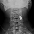
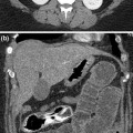
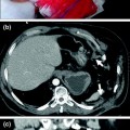
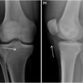
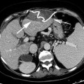
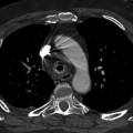
Fig. 10.1
Prosthetic device shifted inside tissues. Migration in the soft tissues of a penile prosthesis consisting of a reservoir shifted in the paravesical region, pumps in the corpus cavernosum, and storage tank in the scrotum (arrow)
So, when the depth of foreign body affects the resolution of US technique, CT-scan or MRI is needed to detect radiolucent foreign body.
10.2 Role of Magnetic Resonance Imaging
Signal intensity artifacts are often encountered during magnetic resonance (MR) imaging [7] (Fig. 10.2), and may be caused by a ferromagnetic foreign body in the imager: these cases are easily identified on radiographs and may limit MRI indication.
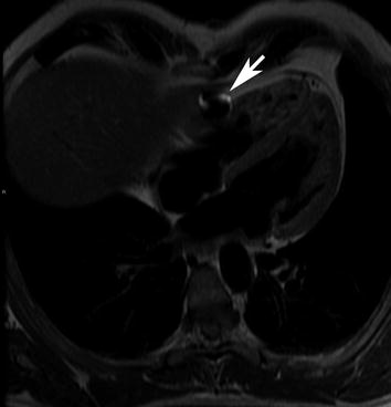

Fig. 10.2
Metallic foreign body occasionally identified by MRI. Cardiac MRI documenting an artifact constant signal (arrow) due to a tiny metal chips at the left ventricle in an underwater fishing man hospitalized under emergency for TIA
The utility of MR imaging in the localization of nonmetallic foreign bodies in the soft tissues of the body is well known [8, 9]. For many time, its high cost has precluded its routine use for evaluating all open injuries suspected of harboring nonradiopaque foreign bodies, however, the recent spread of MRI machines has re-evaluated this topic.
MRI can detect foreign bodies but it may be difficult to distinguish low signal intensity foreign bodies from tendons, scar tissue and calcified area. MRI shows the foreign bodies as low signal or signal void to the muscles on both T1- and T2-weighted images [10]. However, the identification of foreign bodies is difficult on MRI when the bodies are small.
Bodne et al. [11] commented on the absence of signal on MR images of retained foreign bodies composed of wood. Plastic material, which is inert and nonreactive, also can produce signal void on MR images. If the foreign body is long and thin, the lesion may present a “target appearance” when imaged in cross section. More often, MRI is employed to identify infective complications and to differentiate its soft-tissue masses [12, 13], where the detection of these lesions and their extent is better delineated with MRI than CT [11, 14].
When a foreign body is surrounded by inflammatory tissues or a hematoma, a ring of low signal on T1- and high signal on T2-weighted images around the foreign bodies is observed demonstrating a target appearance [15]. The surrounding reactive lesion is easily mistaken for a soft-tissue neoplasm when a foreign body is not identified because the peripheral area of the lesion is strongly enhanced by gadolinium [10]. The central area of the reactive lesion is observed as low intensities on T1- and high intensities on T2-weighted images with no enhancement, which suggests that the lesion is myxomatous or cystic [16, 17


Stay updated, free articles. Join our Telegram channel

Full access? Get Clinical Tree


