George M. Bridgeforth, Thomas Hearty, and Charles Carroll IV
A 81-YEAR-OLD WOMAN FELL DOWN 20 STEPS. SHE COMPLAINS OF MARKED PAIN AND SORENESS WITH AN INABILITY TO MOVE HER WRIST.
CLINICAL PRESENTATION
- Patients may complain of a tear or a “pop.”
- Long-standing cases may present with deformity and/or degenerative arthritis due to carpal instability.
- Clicking may be present with recurrent subluxation during motion.
Clinical Presentation
The scapholunate joint is essential to carpal function. A scapholunate dissociation (SLD), which progresses through several stages, finally ends as a perilunate dislocation. The injury can be completely ligamentous or can be a combined fracture and ligamentous injury. SLD is one of the most common types of carpal instability. Often overlooked, it may lead to weakness and arthritis. Associated with torsion injuries to the wrist with an axial load, SLD is usually caused from a fall on an outstretched hand with the wrist extended, ulnarly deviated, and the forearm pronated.
Patients may exhibit pain and swelling over the proximal carpus near the center of the wrist. Limited range of motion occurs. Clicking from a rotation scaphoid or lunate may be present. To test for wrist stability, the examiner performs a Watson test (scaphoid shift test). The examiner stabilizes the wrist with both hands and places his or her thumb over the volar aspect (palm side) of the scaphoid. While holding the wrist in ulnar deviation and moving it radially, the flexing scaphoid subluxes dorsally, causing a reproducible “click.” There may be a painful “clunk” as the scaphoid reduces back into the radial scaphoid fossa when the thumb pressure is removed. A recurrent click is diagnostic of carpal subluxation. The physician should also check neurovascular status carefully (cold cyanotic fingers with absent or diminished pulses and delayed capillary refill greater than 4 seconds in fingertips).
PATIENT ASSESSMENT 
- Pain, swelling, and soreness over the scaphoid lunate area
- Pain-limited range of motion
- Increased pain with resisted torsion (supination and pronation)
- Possible obvious deformity at the wrist, if there is a perilunate dislocation
- Median nerve dysfunction associated with carpal dislocation
The differential diagnosis includes the following: wrist contusion, radial styloid fracture, scaphoid fracture, scapholunate dislocation, ulnar fracture, triangular fibrocartilage complex (TFCC) tear, perilunate dislocation, and gonococcal septic arthritis, as well as rheumatological conditions and other carpal fractures. It should be noted that SLD is associated with distal radial fractures in more than 50% of cases. Patients with marked pain and swelling of the proximal carpus who do not have any radiological evidence of a distal forearm or wrist fracture may have a TFCC tear. The TFCC, which attaches the ulnar border of the distal radius to the ulna, consists of the radioulnar ligaments, the articular disc, ulnar collateral ligament, the extensor carpi ulnaris tendon, and the meniscus homolog. These structures help stabilize the distal radius and ulna with the neighboring carpals. The mechanism of injury is similar to that of scapholunate dislocations (torsion injuries and falls), and TFCC tears can be misdiagnosed as a more common wrist contusion.
With a TFCC tear, patients can complain of pain, swelling, and tenderness over the distal ulna and proximal wrist. Painful, limited range of motion, especially with torsion (pronation and supination), and particularly with ulnar deviation, may be present. A positive press test—marked wrist pain when the patient tries to use the affected wrist to lift himself or herself up from an examination table or chair—may indicate a TFCC tear. In addition, fractures of the distal ulna, ulnar styloid, or distal radius are associated with TFCC tears.
Radiographic Evaluation
In patients who present with acute swelling, pain, focal tenderness, and limited range of motion, the physician should strongly consider the use of radiographs. It is necessary to take posteroanterior (PA) views with a clenched fist for provocation. Lateral views may also be required. Optional radiographs include a:
- scaphoid view (ulnar deviation): to rule out scaphoid fracture,
- PA with traction, and
- contralateral wrist for comparison.
In addition, a magnetic resonance imaging (MRI) scan or an MRI arthrogram for evaluation of ligament disruption may be helpful.
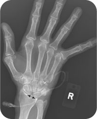
For an SLD, the primary radiographic finding is a 3-mm or greater separation between the scaphoid and the lunate on the PA view. This gap is indicative of a SLD secondary to a torn carpal ligament. It is known as the “Terry Thomas sign,” after the British comedian who had a small gap in his front teeth, and it has also been called the David Letterman sign for similar reasons. Any separation between the carpal bones that is 3 mm or greater is suspicious for a carpal dissociation, and a gap larger than 5 mm is diagnostic (Figs. 39.1 and 39.2). It is important to check a contralateral radiograph to rule out an anatomic variant, and a clenched fist PA view may create the separation not seen on a standard PA view.
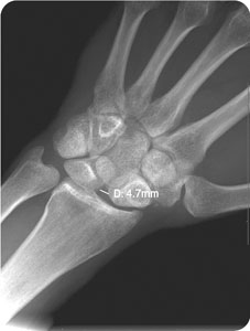
FIGURE 39.1 AP view of the left wrist in a 20-year-old man with left wrist pain after a fall demonstrates widening of the scapholunate space by approximately 5 mm suspicious for scapholunate dissociation.
Related posts:


Stay updated, free articles. Join our Telegram channel

Full access? Get Clinical Tree


