Splenic autoinfarction: Absent or small calcified spleen
Massive splenic infarction: Rare complication defined as when > 50% of spleen is infarcted
Splenic sequestration: Massive splenomegaly
Splenic abscess: Usually due to prior infarcts
• Gallbladder: Gallstones in young patients
• Extramedullary hematopoiesis: Most commonly paravertebral soft tissue masses of homogeneous density
• Kidneys
Papillary necrosis on CT urography
Large kidneys in early phase of disease; gradual atrophy with development of chronic renal failure
• Bones
Widened medullary spaces, thinning of cortex, coarsening of trabecular pattern, and osteopenia
Osteonecrosis and multiple bone infarcts
– High T2WI MR signal in affected areas with serpiginous low T2 signal outline (double-line sign)
H-shaped or “Lincoln log” vertebrae
– Cortical thinning leads to endplate deformities
Osteomyelitis
Persistence of red (cellular) marrow in bones with low-signal marrow on T1WI
– Demand for increased production of RBCs prevents normal conversion of red to yellow marrow
• Findings of iron deposition due to repetitive transfusions
Hyperdense liver on NECT
Liver, spleen, and bone marrow abnormally low signal on all MR pulse sequences (particularly on T2WI)
Involved organs demonstrate characteristic signal loss on in-phase GRE images
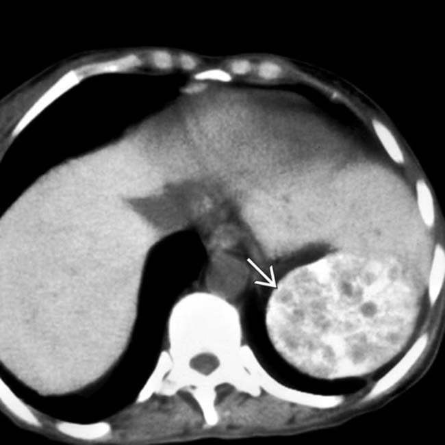
(Left) Axial NECT in a patient who presented with sickle cell anemia and severe left upper quadrant pain demonstrates a heavily calcified and heterogeneous spleen , indicating chronic and possibly acute infarction.
(Right) Splenectomy specimen from the same patient illustrates a mottled spleen with capsular discoloration that was heavily calcified on microscopy.
(Left) Axial NECT in a patient with homozygous sickle cell anemia demonstrates a small and heavily calcified spleen , also known as autosplenectomy.
(Right) Axial CECT in a patient with sickle cell anemia demonstrates multiple wedge-shaped hypodense areas in the spleen, representing massive splenic infarction, an uncommon complication that rarely results in the formation of a splenic abscess.
TERMINOLOGY
Abbreviations
• Sickle cell anemia (SCA)
Definitions
• Inherited hemolytic anemia arising due to abnormal hemoglobin (hemoglobin S), resulting in deformation of red blood cells and leading to microvascular occlusions and infarcts
IMAGING
General Features
• Best diagnostic clue
Small, densely calcified spleen on CT
Papillary necrosis on intravenous pyelogram (IVP) or CT urography
Cholelithiasis in a young African American patient
• Size
Spleen may be undetectable (autosplenectomy) but rarely may enlarge due to sequestration syndrome
CT Findings
• Spleen
Splenic autoinfarction: Absent or small calcified spleen
Massive splenic infarction
– Splenic infarcts usually small and repetitive, leading to autoinfarction
– Massive splenic infarction defined as when > 50% of spleen is infarcted
– Often no precipitating factor, but may be associated with hypoxia (high altitude or mountain climbing)
Splenic abscess: Rare (< 1%) complication usually seen in patients with persistent splenomegaly (rather than autoinfarction) and massive infarcts
Splenic sequestration: Often associated with massive splenomegaly
– Represents severe, life-threatening anemia with massive splenomegaly and reticulocytosis
• Gallbladder: Gallstones in young patients
• Brain: White matter infarcts
• Extramedullary hematopoiesis: May have many different manifestations, including paravertebral soft tissue masses (homogeneous soft tissue density), hepatosplenomegaly, or perinephric soft tissue “rind” encasing kidneys
• Liver: Hyperdense liver due to repeated transfusions


 Liver, spleen, and bone marrow abnormally low signal on all MR pulse sequences (particularly on T2WI)

 , indicating chronic and possibly acute infarction.
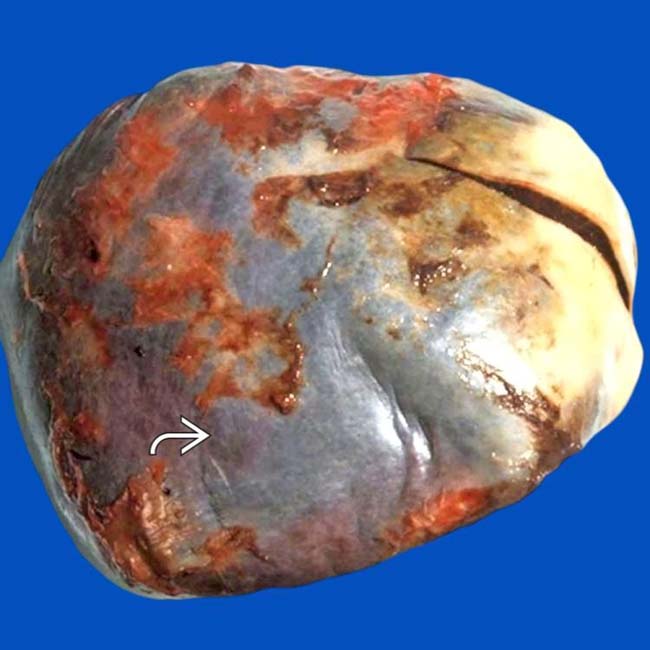
 that was heavily calcified on microscopy.
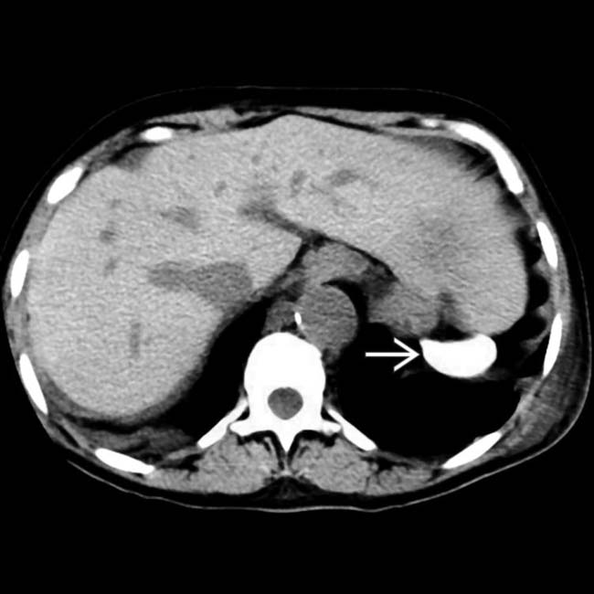
 , also known as autosplenectomy.
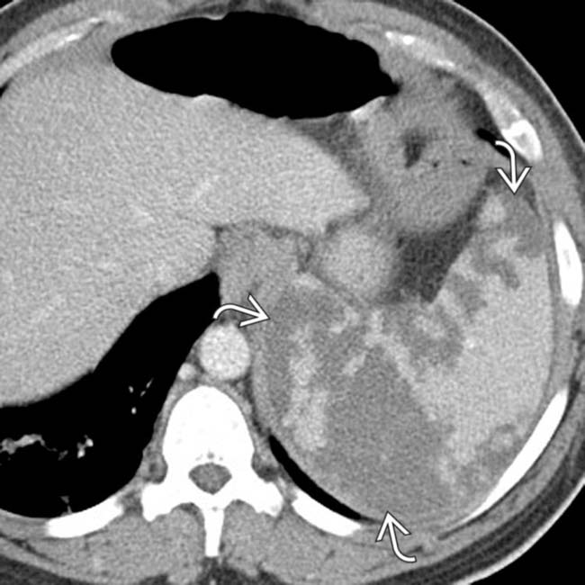
 in the spleen, representing massive splenic infarction, an uncommon complication that rarely results in the formation of a splenic abscess.


 Splenic abscess: Rare (< 1%) complication usually seen in patients with persistent splenomegaly (rather than autoinfarction) and massive infarcts


 Papillary necrosis on CT urography (blunted, irregular calyces, “golf ball-on-tee” appearance, etc.)


 Demand for increased production of RBCs (due to RBC destruction and anemia) prevents normal conversion of red to yellow marrow
 Stimulation of RBC production leads to widened medullary spaces, thinning of cortex, coarsening of trabecular pattern, and osteopenia


 Involved organs demonstrate characteristic signal loss on in-phase GRE images (opposite of steatosis)

 Chest x-ray


