Site of SSTR-positive target lesions
Maximum SUV before PRRNT
Third
Second
First
Liver
S8 apical
12.6
13.2
14.0
S8 apicomedial/central
11.0
14.0
12.2
S2 medial
13.7
15.2
12.9
S5/S8/S7 with central necrosis
16.6
18.1
16.8
S4a
11.6
14.0
12.2
S3
16.6
20.0
13,2
S6 inferolateral
16.2
18.4
15.2
Primary tumor
Ileocoecal region
14.1
16.9
11.6
Iliac bone
Right dorsal
8.0
10.8
–
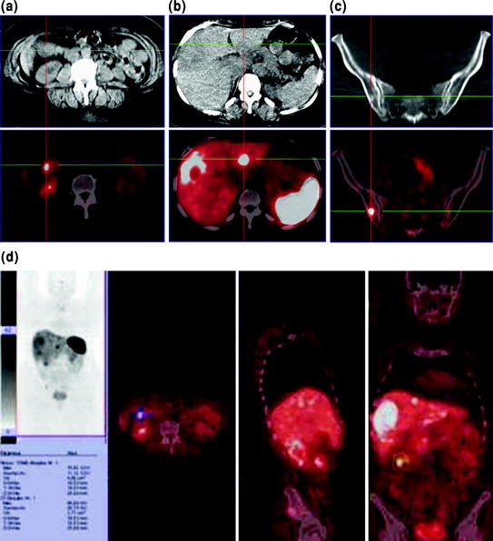
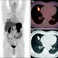
Fig. 1
68Ga-DOTATOC PET/CT showing the following somatostatin receptor-positive lesions: a primary tumor in the ileum (indicated by the cross on image “a” on transversal CT slice (upper row) and on fused PET/CT image (lower row); b in liver segment S3 (indicated by the cross on image “b” on transversal CT slice (upper row) and on fused PET/CT image (lower row); c in the posterior right iliac bone (indicated by the cross on image “c”) on transversal CT slice (upper row), and on fused PET/CT image (lower row); d in the terminal ileum, primary tumor location (maximum-intensity projection, and fused transversal, sagittal, and coronal images)
Abdominal ultrasonography showed multiple focal hyperechogenic lesions in the steatotic liver, the largest being in segment S7 (7.4 cm × 6.7 cm in diameter) and in the left liver lobe with size of 2.3 × 1.9 cm. In the right lower abdomen, a hypoechogenic formation was found with diameter of 1.9 × 2.4 cm.
Kidney scintigraphy performed with 99mTc-MAG3 and tubular extraction rate (TER 219 ml/min, 82% of normal age-adjusted value, lower limit 186 ml/min) were normal and better than the initial value before first PRRNT. GFR (determined by using 99mTc-DTPA) was also normal (83 ml/min, 90% of normal age-adjusted value, lower limit 64 ml/min). Laboratory tests were unremarkable.
18F-fluoride showed one osteoblastic metastasis in the right posterior iliac crest (SUVmax = 21.1) which was also SSTR positive.
Compared with the previous findings, MRI showed regression of liver metastases, particularly the largest ones located in S8 (maximum diameter 65 mm, previously 80 mm) and in S6 (downsized from 38 to 30 mm). Smaller liver metastases also showed regression (16 versus 20 mm).
Chromogranin A was elevated (770 μg/l, cutoff <100 μg/l), as was serotonin in serum (783 μg/l, normal range 40–200 μg/l).
The patient was treated intravenously with 3 GBq of 90Y-DOTATOC and 6 GBq of 177Lu-DOTATOC and tolerated the third PRRNT cycle very well. Whole-body scintigraphy was performed at 23 and 43 h after therapy using the gamma emission of 177Lu (energy window set at 208 keV), and scintigraphic images showed very intense tumor uptake (Fig. 2).
Fig. 2
 via Cation Exchange Cartridges: Versatile Options
 Experience in Peptide Receptor Radionuclide Therapy
 Bombesin Analogs for Receptor-Mediated Imaging
 Novel 68Ga-Labeled Pteroic Acid-Based PET Tracer for Tumor Imaging via the Folate Receptor
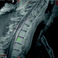 Rare Case of a Large Spinal Meningioma with Mediastinal Extension and Malignant Behavior Classified Histologically as Benign
 Role of 68Ga-Labeled Somatostatin Analogs in the Workup of Patients with NETs: AIIMS Experience


Whole-body scans obtained 43 h after the third cycle of PRRNT using 177Lu- and 90Y-DOTATATE (double-intensity anterior and posterior projections) demonstrating intense somatostatin receptor binding of the therapeutic agent in the primary tumor and in the metastatic lesions in the liver as well as in the small metastasis in the right posterior iliac crest (faintly visible)
Related posts:
via Cation Exchange Cartridges: Versatile Options
Experience in Peptide Receptor Radionuclide Therapy
Bombesin Analogs for Receptor-Mediated Imaging
Novel 68Ga-Labeled Pteroic Acid-Based PET Tracer for Tumor Imaging via the Folate Receptor
Rare Case of a Large Spinal Meningioma with Mediastinal Extension and Malignant Behavior Classified Histologically as Benign
Role of 68Ga-Labeled Somatostatin Analogs in the Workup of Patients with NETs: AIIMS Experience
Stay updated, free articles. Join our Telegram channel
Full access? Get Clinical Tree
