George M. Bridgeforth and George Holmes
A 26-year-old man who slipped on an icy street presents with pain, swelling, and gross deformity of his right ankle.
CLINICAL POINTS
- Torsion may lead to spiral ankle fractures.
- Marked tenderness, pain, and swelling occur at the fracture site.
- Pain-limited ambulation with an inability to take four steps is characteristic.
Clinical Presentation
A spiral fracture is caused by supination of the foot with external rotation force (torsion injuries). Spiral fractures may occur in downhill skiers, who put their feet into rigid boots that fit firmly onto skis. If a ski breaks or changes direction abruptly, an acutely twisted leg may result.
The fracture in the introductory case is known as a Weber B fracture by some orthopedic specialists (see subsequent discussion). It is an oblique fracture of the lateral malleolus that is commonly associated with rupture of the tibiofibular syndesmosis. A more severe fracture is the Maisonneuve fracture (Weber C fracture), a spiral fracture of the distal fibula that extends to the proximal fibula. Fracture of the proximal fibula associated with a torn interosseous ligaments and membrane. These injuries are associated with a disruption of the distal tibiofibular complex.
Pronation is a complex triplanar motion of the foot and ankle consisting of dorsiflexion, ankle eversion, and lateral abduction of the forefoot.* With pronation, the foot and ankle roll outward. Supination is a complex triplanar motion of the foot and ankle consisting of plantar flexion, ankle inversion, and medial adduction of the forefoot. With supination, the foot and ankle roll inward.
Evaluation of the patient is essential. The physician should always check for tenderness of the fibular head and the interosseous region between the distal tibia and fibula and the opposite side for an avulsion fracture of the distal tibia (medial malleolus) or a complete avulsion of the deltoid ligament. Physicians should also inspect the base of the fifth metatarsal for (associated) fractures. In addition, it is always necessary to conduct a thorough neurovascular examination. Signs of pallor, coldness of the foot and ankle (cold dusky limb), diminished or absent dorsalis pedis or posterior tibial pulses, and diminished or absent sensation are important findings. Careful documentation of neurovascular status is essential.
PATIENT ASSESSMENT 
- Marked pain and swelling over the distal fibula
- Pain-limited range of motion
- Limited weight-bearing
The broad-based medial collateral ligament is made up of the deltoid ligament. The lateral collateral ligaments are composed of the anterior and posterior talofibular ligaments and the posterior calcaneofibular. The broad, thick deltoid ligament is stronger than the lateral collateral ligaments. However, if there is a complete avulsion of either the medial or lateral collateral ligaments associated with a (impaction) fracture of the opposite side, the injury is the equivalent of bimalleolar fracture type of injury. From a biomechanical standpoint, there are now two breaks in the structural support on each side of the ankle. Therefore, these injuries are unstable.
Fractures above the tibia plafond (a horizontal line drawn across the distal tibia) are unstable fractures. There is not enough bony stirrup support for ankle stability. These fractures may be associated with tears in the posterior talofibular ligament, which adds to the lack of surrounding structural support.
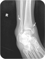
A Tillaux fracture is a fracture of the anterolateral process of the tibia. This injury, which usually occurs in adolescent skate boarders or snow boarders, rarely occurs in adults because the ligaments give way first. It appears as a small avulsion fracture along the lateral border of the tibia and is appreciated best on an oblique view (Fig. 23.1).
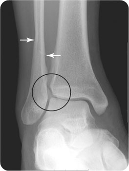
FIGURE 23.1 Tillaux fracture. A 46-year-old man has right ankle pain after playing volleyball. An oblique radiograph demonstrates an acute spiral fracture of the distal right fibula (arrows) and an avulsion of the lateral aspect of the distal tibia (circled).
CLASSIFICATION SYSTEMS
LAUGE–HANSEN CLASSIFICATION
Generally, according to the Lauge–Hansen classification, supination and external rotation are the causes of spiral fractures of the fibula. This classification system resulted from a cadaver study conducted in 1950. During this study, scientists applied a directional force either rotational (internal vs. external) or linear (abduction vs. adduction) to a cadaver ankle placed in either supination or pronation. They made an attempt to classify the resulting fracture patterns. Although this complex classification has limited clinical applications, it is a landmark biomechanical study of ankle fractures.
WEBER CLASSIFICATION
- Type A fractures: These are transverse avulsion (or chip) fractures of the distal fibula. Weber A fractures occur below the level of the tibial plafond. Therefore, these are stable fractures in which the deltoid ligament, the distal tibiofibular complex, and the medial malleolus are intact. However, if the medial malleolus is fractured, then these injuries can be unstable. Generally, these are inversion injuries (see Fig. 23.2).
- Type B fractures: These are oblique fractures of the distal fibula. Weber B fractures are caused by an external rotation force to a supinated foot. They usually occur at the level of the distal tibiofibular complex. They may be associated with an avulsion chip fracture, a transverse fracture of the medial malleolus, or a disrupted deltoid ligament (increased in the tibiotalar space). An associated fracture of the medial malleolus or a disrupted deltoid ligament constitutes a bimalleolar fracture. If there is an associated third fracture of the posterior lip of the tibia, then the injury is a trimalleolar fracture. Weber B (oblique) fractures are above the tibial plafond and should be considered unstable fracture unless proven otherwise (see Fig. 23.3).
- Type C fractures: These are spiral fractures of the distal fibula. They are unstable fractures that extend above the level of the tibial plafond. The external rotation force applied to a supinated foot causes a disruption of distal tibiofibular complex. Moreover, it is associated with a disruption of the distal tibiofibular complex. The spiral fracture of the fibula may extend to the upper fibula (Maisonneuve fracture) (Fig. 23.4). These fractures may be associated with fractures of the distal tibia or disruptions of the deltoid ligament (bimalleolar fractures). If there is an associated third fracture involving the posterior lip of the tibia, then the injury is a trimalleolar fracture.
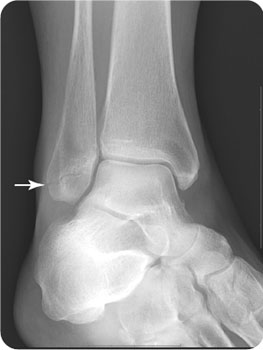
FIGURE 23.2 A 62-year-old woman who fell presents with lateral right ankle pain. The oblique radiograph demonstrates a transverse fracture of the right distal tibia.
Related posts:


Stay updated, free articles. Join our Telegram channel

Full access? Get Clinical Tree


