Perisplenic hematoma: Located adjacent to spleen and implies disruption or rupture of splenic capsule
Intraparenchymal hematoma: Typically round or irregular in shape
Subcapsular hematoma: Constrained by splenic capsule and crescentic in shape
• Sentinel clot sign: Highest density blood localizes adjacent to spleen (or any site of injury)
Indicates splenic injury even without demonstrable laceration
• Parenchymal laceration: Irregular linear, branching, or stellate area of nonenhancing low attenuation
• Splenic fracture: Deep laceration extending from outer capsule through splenic hilum
• Splenic infarction: Unusual (< 2% of cases) in the setting of trauma, and can be segmental or complete
• Active arterial extravasation: High-attenuation focus isodense with aorta, surrounded by lower attenuation clot or hematoma
Distinction between active extravasation and pseudoaneurysm using delayed phase images
TOP DIFFERENTIAL DIAGNOSES
• Splenic cleft
• Splenic abscess
• Splenic infarct
• Splenic cyst
• Lymphoma and splenic tumors
CLINICAL ISSUES
• Most commonly injured solid abdominal organ in blunt trauma and most common abdominal organ injury requiring surgery
• Prone to develop delayed hemorrhage, but excellent prognosis with early intervention (surgery/embolization)
• Identification of active arterial extravasation or pseudoaneurysm best predictor of need for surgery and failure of nonoperative management
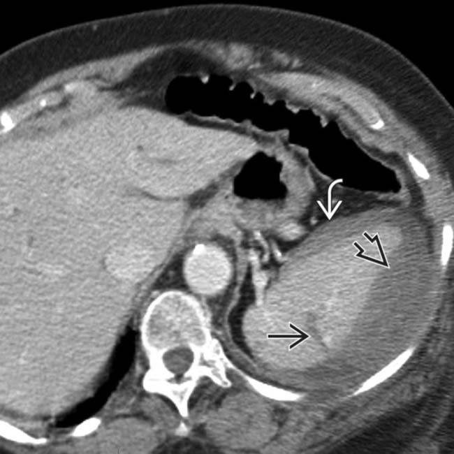
(Left) Axial CECT in an 87-year-old woman who fell at a nursing home demonstrates a splenic parenchymal laceration and intraperitoneal blood , as well as a lentiform heterogeneous and higher attenuation collection flattening the normal convex lateral splenic contour, representing a subcapsular hematoma .
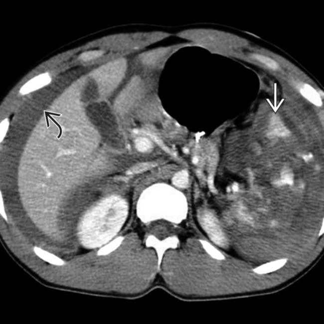
(Right) Axial CECT in a 23-year-old man injured in a motor vehicle accident shows a shattered spleen with a sentinel clot in the perisplenic region and large hemoperitoneum .
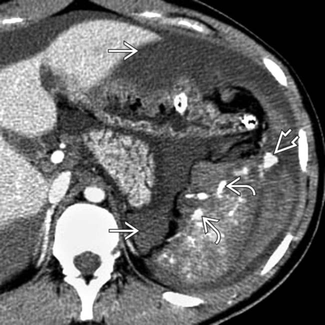
(Left) Axial CECT in a 19-year-old man who was an unrestrained passenger in a motor vehicle accident shows marked upper abdominal hemoperitoneum , a shattered spleen with intrasplenic high-attenuation pseudoaneurysms , and a focus of active arterial extravasation lateral to the spleen within the peritoneal cavity .
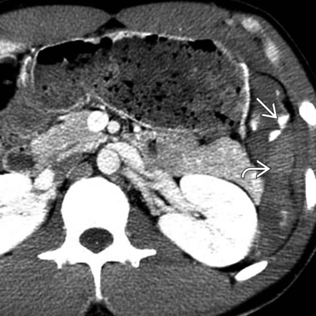
(Right) Axial CECT in the same patient shows the active arterial extravasation extending into the left paracolic gutter with surrounding hemoperitoneum .
TERMINOLOGY
Synonyms
• Splenic laceration or splenic fracture
Definitions
• Splenic parenchymal injury ± capsule disruption
IMAGING
General Features
• Best diagnostic clue
Low-attenuation splenic laceration with high-density active bleeding
• Morphology
Lacerations: Linear or jagged edges
Fracture: Laceration extending from outer cortex to hilum
Subcapsular hematoma: Flattened contour of splenic parenchyma
Radiographic Findings
• Radiography
Abdominal radiography
– Left upper quadrant soft tissue mass
– Signs of intraperitoneal fluid with widening of distance between flank strip and descending colon
– Fluid in pelvis with prominent pelvic “dog ears”
– Left rib fractures, pneumothorax, pleural effusion
CT Findings
• NECT
High-attenuation hemoperitoneum > 30 HU or perisplenic clot > 45 HU
– Perisplenic, intraparenchymal, or subcapsular hematoma
Perisplenic hematoma: Located adjacent to spleen and implies disruption or rupture of splenic capsule
Intraparenchymal hematoma: Typically round, ovoid, or irregular in shape
Subcapsular hematoma: Constrained by splenic capsule; crescentic in shape and compresses lateral margin of parenchyma
Sentinel clot sign: Highest density blood localizes adjacent to spleen (or any site of injury)
– Indicates splenic injury even in absence of demonstrable laceration
Layered or lamellated clot if bleeding is intermittent
• CECT
Parenchymal laceration: Irregular linear, branching, or stellate area of nonenhancing low attenuation within parenchyma
– May extend to splenic capsule resulting in capsular tear
– Should become less conspicuous on follow-up imaging
Splenic fracture: Deep laceration extending from outer capsule through splenic hilum
Splenic infarction: Unusual (< 2% of cases) in setting of trauma
– Can be segmental or complete
– Wedge-shaped area of hypoattenuation
– Due to arterial thrombosis after intimal injury
– Risk of delayed rupture or abscess formation
Active arterial extravasation: High-attenuation focus isodense with aorta, surrounded by lower attenuation clot or hematoma
– May be linear (spurting vessel) or rounded (pseudoaneurysm): Distinction is made using delayed phase images
Active extravasation (unlike pseudoaneurysm) changes in size and morphology between initial and delayed phases
Although delayed images are not routinely included in most trauma protocols, addition of delayed images can be helpful if there is site of suspicion noted on initially acquired portal venous phase images
Ultrasonographic Findings
• Subtle laceration may be missed, as ultrasound is insensitive for parenchymal injury
Lacerations can be hypoechoic or isoechoic to splenic parenchyma and can be very difficult to detect with US
• Free intraperitoneal fluid with low-level echoes representing hemoperitoneum and echogenic perisplenic clot
• Hematoma should be avascular
Angiographic Findings
• Avascular parenchymal laceration with amorphous parenchymal extravasation
• Flattened lateral contour of spleen due to subcapsular hematoma
• Rounded contrast collections (pseudoaneurysms)
Imaging Recommendations
• Best imaging tool
CECT
• Protocol advice
Arterial phase images more sensitive for active extravasation or pseudoaneurysm
Portal venous phase images more sensitive for parenchymal injury (i.e., laceration)
Only gold members can continue reading. Log In or Register to continue


 and intraperitoneal blood
  , as well as a lentiform heterogeneous and higher attenuation collection flattening the normal convex lateral splenic contour, representing a subcapsular hematoma
  .
.
 in the perisplenic region and large hemoperitoneum
  .
.
 , a shattered spleen with intrasplenic high-attenuation pseudoaneurysms
  , and a focus of active arterial extravasation lateral to the spleen within the peritoneal cavity
  .
.
 extending into the left paracolic gutter with surrounding hemoperitoneum
  .
.


 High-attenuation hemoperitoneum > 30 HU or perisplenic clot > 45 HU


 Parenchymal laceration: Irregular linear, branching, or stellate area of nonenhancing low attenuation within parenchyma
 Active arterial extravasation: High-attenuation focus isodense with aorta, surrounded by lower attenuation clot or hematoma


