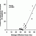
Fig. 1
Comparison of papillary thyroid microcarcinoma (PTMC) according to tumour size: In 349 patients with PTCM the tumour size was significantly correlated with the prevalence of lymph node metastasis (p < 0.01)
4 Histology
Thyroid microcarcinomas are most often papillary (65–99 %) and only rarely follicular (0.3–23.6 %) tumours (Baudin et al. 1998; Lee et al. 2006). The wide range in prevalence of the follicular microcarcinoma may at least in part be due to its substantial similarity with the follicular variant of PTMC. However, both patients with follicular and those with papillary microcarcinoma have an excellent prognosis (Rahbar et al. 2008). At the Department of Nuclear Medicine of the University of Münster, a tertiary referral centre for differentiated thyroid cancer, thyroid microcarcinoma showed excellent event-free survival rates irrespective of their histology (Fig. 2). Accordingly, the European guidelines recommend common therapeutic algorithms for both types of differentiated microcarcinomas (Pacini et al. 2006). The PTMC is often non-encapsulated and sclerosing, although encapsulated variants exist. The tumour is commonly located near the thyroid capsule. Small PTMC <1 mm frequently present with a follicular architecture and lack stromal sclerosis (DeLellis et al. 2004). In contrast, PTMC with a mean diameter of about 2 mm often show a prominent desmoplastic stroma. Larger tumours with an average size of 5 mm are less common and often characterised by a papillary pattern. The more virulent variants of PTMC such as tall cell, columnar cell, oncocytic and diffuse sclerosing subtypes are rare. The prevalence of oncocytic and tall cell variants in PTMC is only 0.8 % (Pelizzo et al. 2004). However, the corresponding value for the sclerosing subtype amounts to 5.0 % (Pelizzo et al. 2004). Until 2009, the American and European guidelines concordantly recommended radioiodine ablation after total or near-total thyroidectomy in these more aggressive subtypes of PTMC (Cooper et al. 2006; Luster et al. 2008). In contrast, the current American guidelines have confined the use of radioiodine ablation to PTMC patients with extrathyroidal extension and lymph node or distant metastasis, respectively (Cooper et al. 2009). However, this highly restrictive approach is not shared by European stakeholders who still suggest radioiodine ablation in selected PTMC patients with T1 N0 M0 (Table 1).
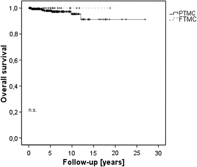

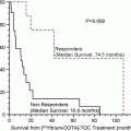
Fig. 2
Event-free survival of patients with thyroid microcarcinoma of papillary (PTMC) and follicular histology (FTMC). There was no significant difference between the event rates in patients with PTMC and FTMC (p = n.s)
Table 1
Summary of the international guidelines on the indication of radioiodine ablation in patients with papillary thyroid microcarcinoma
ATA | ETA | EANM |
|---|---|---|
No patients with T1 N0 M0 | Selected patients with T1 N0 M0 multifocality unfavourable histology incomplete surgery | Selected patients with T1 N0 M0 multifocality unfavourable histology history of radiation exposure |
Patients with T3 or N1/M1-categories | Patients with T3, R1 or N1/M1-categories; relative indication for <18 years | Patients with T3 or N1/M1-categories |
5 Molecular Characteristics of PTMC
From a nuclear medicine perspective, the deeper insights into the molecular characteristics of PTMC will have a growing impact on the therapeutic management of these patients. Several genetic alterations have been described in PTMC which are associated with distinct biological features. Most common mutations in papillary carcinoma are point mutations of the BRAF and RAS genes and receptor tyrosine kinase (RET) rearrangements, all of which encode proteins that act along the same intracellular signalling pathway leading to the activation of the mitogen-activated protein kinase (MAPK) cascade (Nikiforov 2008). Chromosomal rearrangements of the RET gene represent the most common structural genetic alterations in papillary carcinoma. These rearrangements have been found in up to 52 % of PTMC and represent an early event in the process of thyroid cell transformation (Adeniran et al. 2006; Viglietto et al. 1995). More aggressive PTMCs which present with cervical lymph node metastases often show loss of the p27 gene (Khoo et al. 2002). In addition, cyclin D1 showed significantly higher median expression in PTMC with metastases compared to those without, indicating a correlation to tumour aggressiveness (Londero et al. 2008). Lim et al. (2007) have shown that the absence of epidermal growth factor receptor expression is closely correlated with lymph node metastasis and extrathyroidal growth. Furthermore, they have demonstrated that the absence of cyclooxygenase-2 expression is associated with multiplicity and bilaterality.
6 Therapy
The guidelines of the ATA, ETA and the Therapy Committee of the EANM agree on a risk-adapted therapeutic management of patients with PTMC as outlined below (Pacini et al. 2006; Cooper et al. 2006a, b; Luster et al. 2008).
6.1 Surgery
The optimal surgical treatment of PTMC is still debatable (Roti et al. 2006; Besic et al. 2008). In a meta-analysis, Roti et al. have found that total/near-total thyroidectomy is performed in 72 %, subtotal thyroidectomy in 11 % and lobectomy in 17 % of cases (Roti et al. 2008). According to Ito et al. therapeutic lymph node dissection is carried out in about 10 %, whereas prophylactic lymph node excision is performed in about 56 % (Ito et al. 2004).
The European guidelines recommend distinct approaches depending on the method of discovery and the risk stratification of the PTMC:
(a)
When PTMC is diagnosed after lobectomy for benign thyroid disease completion thyroidectomy should be performed in case of the following risk factors:
(I)
columnar cell variant
(II)
tall cell variant
(III)
diffuse sclerosing variant
(IV)
multifocality
(V)
extrathyroidal growth
(VI)
locoregional metastasis
(VII)
distant metastasis
(b)
In case of a preoperative diagnosis of the PTMC by fine-needle aspiration cytology standard (near-)total thyroidectomy should be performed. This procedure allows the elimination of multifocal disease and decreases recurrence rates.
(c)
If the PTMC presents with locoregional or distant metastases routine total or near-total thyroidectomy with lymph node dissection is recommended.
The two major complications of thyroid surgery are permanent post-operative recurrent laryngeal nerve palsy and hypocalcaemia. According to the prospective German Thyroid Multicentre Study performed 1998 through 2001, Dralle et al. have found complication rates of only 1.7 % for permanent recurrent laryngeal nerve palsy per nerves at risk and 6.8 % for permanent hypocalcaemia (Dralle et al. 2004). The complication rates depend on the indication and extent of surgery as well as the specialisation of the surgeons (Dralle and Sekulla 2005). Thus, in centres of endocrine surgery the complication rates of permanent hypocalcaemia and recurrent laryngeal nerve palsy are significantly lower than those in low-volume hospitals. In our tertiary referral centre the rates of transient and/or permanent unilateral recurrent laryngeal nerve palsy vary between 1 and 15 % and are inversely proportional to the experience of the surgeon.
6.2 Radioiodine Ablation
The relatively low prevalence and lengthy overall survival of patients with differentiated thyroid cancer and, particularly, PTMC constrain the execution of large-scale prospective studies. Therefore, the level of evidence with regard to the therapeutic management of these patients is low in many instances. Accordingly, the authors of the EANM guidelines relied significantly on their clinical experience to supplement the observations reported in the literature (Luster et al. 2008). Thereby, the current EANM guidelines provide clear and comprehensive therapeutic algorithms of high relevance to everyday practice. This is of particular importance for the adequate therapeutic management of patients with PTMC and diverse risk-profiles.
According to the results of a meta-analysis only about 17 % of patients with PTMC undergo radioiodine ablation (Roti et al. 2008). Chow et al. have found a lower rate of lymph node recurrence after radioiodine ablation in patients with initially lymph node-negative PTMC (Chow et al. 2003). However, these findings could not be confirmed (Baudin et al. 1998). Most PTMC are diagnosed incidentally after surgery for benign thyroid disease and lack the risk factors mentioned above (Pazaitou-Panayiotou et al. 2007). However, in selected patients with PTMC at higher risk (unfavourable histology, multifocality, extrathyroidal growth or metastasis) post-surgical radioiodine ablation is definitively indicated (Table 1).
In addition, the Therapy Committee of the EANM has defined further parameters which should be considered when deciding whether to perform radioiodine ablation in patients with PTMC:
(I)
positive family history
(II)
history of neck radiation exposure
(III)
larger tumour size
(IV)
closeness of the tumour to the thyroid capsule
(V)
vascular invasion
(VI)
cancer-related genetic findings (in the future).
In these cases, the decision on the post-operative radioiodine treatment should be made after individual discussions of the pros and cons with each patient. In general, radioiodine ablation has only minor side effects, such as sialadenitis, xerostomia or nausea (Dietlein et al. 2007). In addition, the patient′s needs of security or possible fears of radiation should be addressed.
6.3 Thyrotropin-Suppressive Therapy
The American and European guidelines recommend a differential L-thyroxine therapy according to the risk profile of the patients with PTMC mentioned above (Pacini et al. 2006; Cooper et al. 2009). In low-risk patients, L-thyroxine replacement therapy is adequate. In these cases Schlumberger et al. recommend to aim for a serum TSH level within the lower part of the normal range (0.5–1.0 mU/l) (Schlumberger et al. 2004). In high-risk PTMC patients with apparent remission after treatment TSH-suppressive therapy (serum TSH ≤ 0.1 mU/l) may be persued for 3–5 years in order to inhibit the TSH-dependent growth of residual cancer cells (Cooper et al. 1998; McGriff et al. 2002). In high-risk patients considered in complete remission therapy can be shifted from suppressive to replacement (Pazaitou-Panayiotou et al. 2007).
Stay updated, free articles. Join our Telegram channel

Full access? Get Clinical Tree


