Fig. 39.1
Injection points of ICG solution
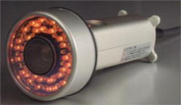
Fig. 39.2
PDE camera (PDE-neo)
We observed the normal and abnormal lymph flows in real time, but it was difficult to observe the entire images of lymph flows in the upper limbs (Fig. 39.3) due to the wide observation field. Therefore, we traced the IF images directly on the skin of the subjects’ arm with a black felt pen (Fig. 39.4).
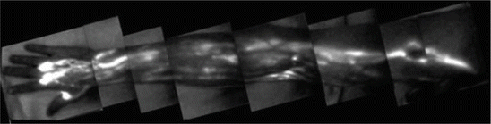
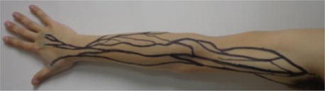
Fig. 39.3
Entire images of lymph flow of upper limb
Fig. 39.4
Tracing of IF images by black felt pen
39.3 Results
39.3.1 The Normal IF Lymphography of Superficial Lymph Flows
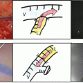
We were able to observe several IF images in which the ICG solution ran from the forearm to the upper arm immediately after the injection of the solution in all five healthy volunteers. We also observed that the IF images reached the axilla area within 4–5 min after the injection of ICG in the normal arms. When the ICG solution was injected at the base of the thumb and forefinger, the lymph vessels were observed to run nearly straight to the deltoid muscle area. When the ICG solution was injected at the root of the third finger, the lymph vessel’s lines ran toward the axilla region after surrounding the elbow. The networks are demonstrated in an upper arm and forearm in Fig. 39.5.
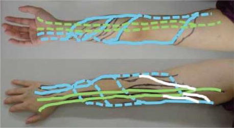
Fig. 39.5
Green lines: the lymph flows on the radial side. Blue lines: the lymph flows on the ulnar side. White lines: these networks in an upper arm and forearm
39.3.2 Characteristic ICG Fluorescence Images of BCRL Patients
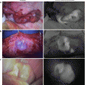
The speed of the IFG lymphatic flow became extremely slow compared to the normal arms. The normal IFG lymph flows were observed as a linear pattern (Fig. 39.6a). Abnormal IF images of lymphedema were classified into three characteristic patterns called “dermal backflow (DB)”: the splash (Fig. 39.6b), stardust (Fig. 39.6c), and diffuse (Fig. 39.6d) patterns. In the present study, these patterns were confirmed in the BCRL patients with lymphedema of the limbs.
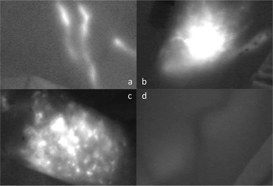
Fig. 39.6
Characteristic patterns of ICG fluorescence images. (a): Linear (normal lymph flow), (b): splash, (c): stardust, and (d): diffuse. Panels (b–d) show an abnormal pattern of BCRL called dermal backflow (DB)
39.3.3 Evaluation of the Effect of CDP for the Upper Limbs Using the IF Method
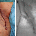
A case was reported in which the lymphedema of the upper limb was dramatically improved by CDP (Figs. 39.7 and 39.8). We observed many instances of DB (the splash pattern) in the IF images of the superficial lymph flow in the BCRL patients’ forearms before CDP (Fig. 39.7a). We marked the lymphedema points for the IF imaging (Fig. 39.7b). The splash pattern decreased and the linear pattern appeared on the forearm in one patient after 1 year of CDP treatment (Fig. 39.8a, b).
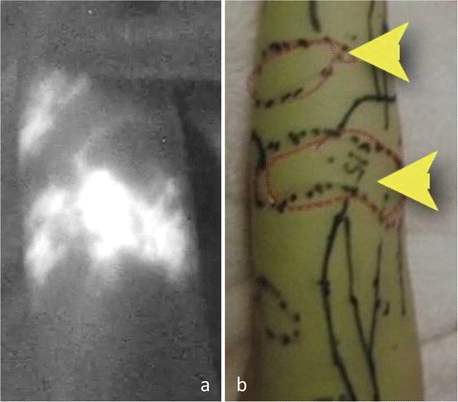
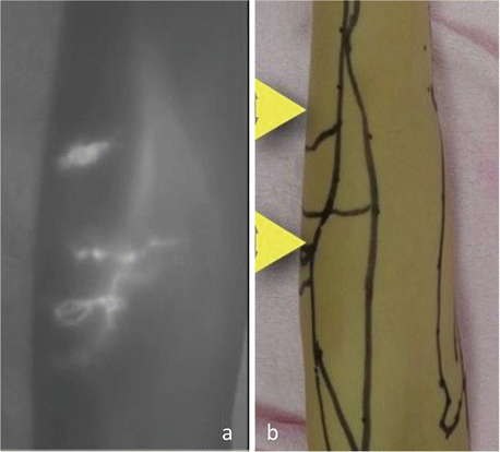
Fig. 39.7
IF image (a) and marking image (b) before CDP. Arrowheads: the splash pattern
Fig. 39.8
IFG image (a) and marking image (b) after CDP. Arrowheads: the improved points
39.4 Discussion
The incidence of BCRL has a strong correlation with axillary dissection and radiotherapy [9–12]. BCRL is major complication that interferes with a patient’s quality of life, and it induces psychological stress. The effectiveness of CDP for BCRL patients has been shown [1, 2].
Related posts:
 ICG Fluorescent Image-Guided Surgery in Head and Neck Cancer
 Regional Lymph Node Dissection Assisted by Indocyanine Green Fluorescence Lymphography and Angiography for Stage III Melanoma
 Fluorescence Imaging for Intraoperative Identification of Pancreatic Leak
 Intraoperative Detection of Hepatocellular Carcinoma Using Indocyanine Green Fluorescence Imaging
 Indocyanine Green Fluorescent Lymphography and Microsurgical Lymphaticovenous Anastomosis
 Detection of Hepatic Micrometastases from Pancreatic Cancer
Stay updated, free articles. Join our Telegram channel

Full access? Get Clinical Tree


