Fig. 11.1
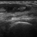
Flexor tendons at the level of the proximal phalanx. The flexor tendons at the level of the proximal phalanx are oriented differently, so the degree of anisotropy they present varies with the inclination of the transducer
The extensor tendons run along the dorsal aspects of the digits. Like the flexor tendons, they are anatomically complex. Distally, each divides to form three laminae, which insert into the base of the middle phalanx (the middle lamina) and the base of the distal phalanx (the two lateral laminae). Of the structures that stabilize these tendons, one of the most important is a retinaculum known as the sagittal band, which located at the level of the metacarpophalangeal joint [5].
Tears and tenosynovitis are the lesions most commonly encountered in the digital tendons of the hand [6].
Tears are usually caused by acute trauma (cuts) (Figs. 11.2, 11.3, 11.4), but they can also be associated with rheumatoid arthritis or blunt trauma [6, 7]. They are more common at the level of the extensor tendons and are classified as partial (Figs. 11.2, 11.3) or complete (Fig. 11.4). Complete, full-thickness tears are characterized by the absence of the tendon (Fig. 11.4) and the presence of a hematoma in the acute phase. Partial tears are manifested by incomplete loss of tendon continuity with thickening and hypoechogenicity at the level of the lesion (Figs. 11.2, 11.3) and (in the acute phase) blood within the tendon sheath. From a functional point of view, this category of lesions also includes avulsion fractures, which are manifested by the presence of a detached bone fragment (Fig. 11.5).

Get Clinical Tree app for offline access
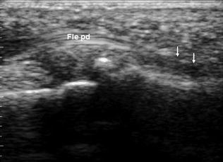
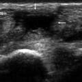
Fig. 11.2
Acute partial tear of a flexor digitorum profundus tendon (Fle pd). The sonogram reveals incomplete loss of tendon continuity, thickening, and hypoechogenicity at the level of injury. A moderate amount of fluid can also be appreciated within the tendon sheath
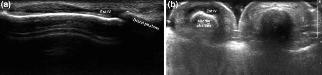
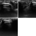
Fig. 11.3
Chronic partial tear of the extensor tendon of the ring finger (Est IV). Sonography reveals a thickened, hypoechoic tendon at the level of the middle phalanx (a long-axis scan, b short-axis scan)
Fig. 11.4
Complete tear of the flexor pollicis longus tendon. Sonography (a) demonstrates absence of the tendon at the level of the interphalangeal joint (confirmed by magnetic resonance imaging) (b) and retraction and deflection of the tendon stumps (a, c) (arrows)
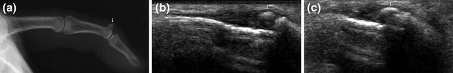
Fig. 11.5
Hammer finger. The radiographic examination reveals (a) an avulsed bone fragment (arrow) and obligatory flexion of the distal phalanx. Sonography (b long-axis view, c


Related posts:
Stay updated, free articles. Join our Telegram channel
Full access? Get Clinical Tree
