The Abdominal Wall and Diaphragm


The Abdominal Wall and Diaphragm


Sharlette Anderson


The body is divided into two major cavities: the dorsal cavity and the ventral cavity. The dorsal, or posterior, cavity is completely encased in bone and is subdivided into the cranial cavity, which houses the brain, and the spinal cavity, which houses the spinal cord. The ventral cavity is divided by the diaphragm into the thoracic cavity superiorly and the abdominopelvic cavity inferiorly. The abdominopelvic cavity is subdivided into the abdomen and the pelvis though no physical barrier separates them. The abdominopelvic cavity is enclosed by the abdominal wall. This chapter focuses on the abdominal wall and diaphragm.

Sonography of the abdominal wall is an efficient and effective way of evaluating the integrity, structure, and function of the abdominal wall and diaphragm. Real-time imaging facilitates the evaluation of muscle contraction and relaxation as well as the motion of contents of the abdominal cavity. The ability to document motion helps sonographers demonstrate the motion of hernias through the layers of the abdominal wall, the changes in muscle fiber motion secondary to trauma or mass, and the changes in diaphragm motion related to paralysis or inflammation.

Sonography is a safe, cost-effective, and widely available method of imaging for most patients. Its portability and lack of radiation exposure support serial evaluation to monitor the progression of pathology and response to therapeutic measures. Although the transmission of any type of energy into the body has the potential to cause bioeffects, the benefits of sonographic evaluation of the abdominal wall and diaphragm by a competent sonographer far outweigh the risks, which, according to current literature, are negligible.1,2


EMBRYOLOGY

In the fourth week of development, the embryo folds from a flat disk into a tubular structure. The caudal end of the embryo folds cranially, moving the connecting stalk from the tail region of the embryo to the ventral surface, forming the site of the future umbilical cord insertion. At the same time, the sides of the disk fold ventrally, folding in part of the yolk sac to form the gut tube. This process also forms the ventral body cavity and the body walls (Fig. 4-1).

Diaphragmatic development is a complicated coordination of muscle, connective tissue, vessels, and nerves. Initially, the septum transversum develops to form a separation between the thoracic and abdominal cavities. It becomes anchored to the anterior wall of the embryo between the
heart and the liver. Paired pleuroperitoneal folds then extend medially from the lateral portions of mesoderm (the middle germ layer of the embryo) toward the septum transversum to form a scaffold for muscle fibers and connective tissue to form the diaphragm and its central tendon3 (Fig. 4-2).


REGIONS AND QUADRANTS

To provide a more standardized way of describing the location of organs, pain, or pathology, the abdominopelvic cavity is divided into four quadrants by a midline vertical line extending from the xyphoid process to the symphysis pubis and a horizontal line through the umbilicus. The four quadrants are as follows: (1) right upper quadrant (RUQ), (2) left upper quadrant (LUQ), (3) left lower quadrant (LLQ), and (4) right lower quadrant (RLQ). For more specific descriptions, the abdomen is further divided into nine regions by dividing it with two vertical lines at the right and left midclavicular planes and two horizontal lines at the inferior costal margin and the level of the fifth lumbar vertebra and iliac tubercles. The nine regions are as follows: (1) right hypochondrium, (2) epigastrium, (3) left hypochondrium, (4) right lumbar, (5) umbilical, (6) left lumbar, (7) right iliac fossa, (8) hypogastrium, and (9) left iliac fossa4,5 (Fig. 4-3).


ANATOMY

There are no physical divisions of the abdominal wall; it is a continuous structure. However, for descriptive purposes, it is divided into the anterior, right and left lateral, and posterior walls. The boundary between the anterior and lateral walls is indefinite, so clinicians often discuss the anterolateral abdominal wall as a single unit4,5 (Fig. 4-4).


Anterolateral Abdominal Wall

The anterolateral wall extends from the thoracic cage to the pelvis. Superiorly, it is bounded by the cartilages of the 7th to 10th ribs and the xiphoid process. Inferiorly, it is bounded by the inguinal ligament and iliac crests, pubic crests, and pubic symphysis of the pelvic bones.6,7


Layers

When describing abdominal wall anatomy, it is important to distinguish between fascia and aponeuroses. A fascia is a fibrous tissue network located between the skin and the underlying structures. It is richly supplied with both blood vessels and nerves. The fascia is composed of two layers: a superficial layer and a deep layer. The superficial fascia is attached to the skin and is composed of connective tissue containing varying quantities of fat. The deep fascia is loosely connected to the superficial fascia by fibrous strands. The deep fascia covers the muscles and partitions them into groups. Although the deep fascia is thin, it is more densely packed and is stronger than the superficial fascia; however, neither the superficial fascia nor the deep fascia possesses any notable internal strength because they are a condensation of connective tissue organized into definable homogeneous layers within the body.7,8

Aponeuroses are layers of flat tendinous fibrous sheets fused with strong connective tissue that attach muscles to fixed points, functioning like a tendon, so they are quite strong. Aponeuroses have minimal vascularity and innervation. The abdominal wall aponeuroses are primarily located in the ventral abdominal regions with a primary function to join muscles to the body parts that the muscles act upon. The most readily known abdominal aponeurosis is the rectus sheath.
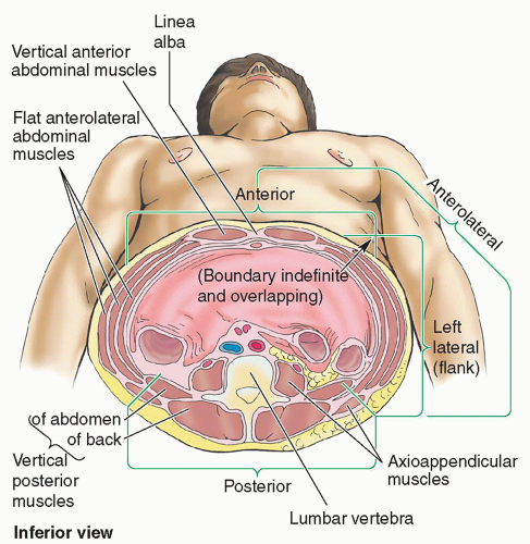
7,8

The abdominal wall appears as a laminated structure when viewed in cross section.7 From superficial to deep, the layers include the following: (1) skin, (2) subcutaneous tissue (superficial fascia), (3) muscles and their aponeuroses, (4) deep fascia, (5) extraperitoneal fat, and (6) the parietal peritoneum.4,6,7 The skin attaches loosely to most of the subcutaneous tissue except at the umbilicus where the attachment is firm.4,8

The subcutaneous tissue anterior to the muscle layers makes up the superficial fascia. Superior to the umbilicus,
it is consistent with that found in most regions. In contrast, inferior to the umbilicus, the deepest part of the subcutaneous tissue is reinforced with elastic and collagen fibers and is divided into two layers. The first is a superficial fatty layer (Camper fascia) containing small vessels and nerves. Camper fascia gives the body wall its rounded appearance. The second layer is a deep membranous layer (Scarpa fascia) and it consists of a combination of fat and fibrous tissue that blends with the deep fascia.4,8 The membranous layer continues into the perineal region as the superficial perineal fascia (Colles fascia)4 (Fig. 4-5).


The three anterolateral abdominal muscle layers and their aponeuroses are covered by superficial, intermediate, and deep layers of extremely thin investing fascia.
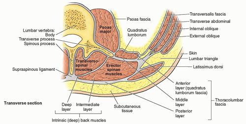
4,8 The investing layer of fascia is located on the external aspects of the three muscle layers and is not easily separated from the external muscle layer, the epimysium. Varying thicknesses of membranous and areolar sheets of endoabdominal fascia line the internal aspects of the wall. Although the endoabdominal fascia is continuous, different regions are named based on the muscle or aponeurosis it lines. For example, the portion lining the deep surface of the transversus abdominis muscle and its aponeurosis is the transversalis fascia. Internal to the endoabdominal fascia is the parietal peritoneum. The distance separating the parietal peritoneum from the endoabdominal fascia is determined by the variable amounts of extraperitoneal fat in the fascia.4 The parietal peritoneum is the outer layer of the serous membrane lining the abdominopelvic cavity formed by a single layer of epithelial cells and supporting connective tissue4 (see Fig. 4-5).


Muscles

There are five bilaterally paired muscles in the anterolateral abdominal wall and one unpaired muscle (Table 4-1). Located bilaterally on the anterior abdominal wall are the rectus abdominis muscles (see Fig. 4-4). The rectus abdominis is a long, broad, vertical, strap-like muscle that is mostly enclosed in the rectus sheath. Also located on the
anterior abdominal wall in the rectus sheath is the pyramidalis muscle. The pyramidalis, a small triangular muscle, extends from its base, originating on the pubic bone to its apex inserting into the midline linea alba approximately halfway between the pubic symphysis and the umbilicus. This muscle lies deep to the anterior rectus fascia and superficial to the rectus abdominus muscle. The pyramidalis muscle is present in about 80% to 90% of people and can be easily harvested for use in reconstructive surgery when needed. Its function is poorly understood, but it helps to maintain tension on the linea alba, supporting the tone of the anterior abdominal wall4,6,9 (Fig. 4-6).


There are three flat, bilaterally paired muscles of the anterolateral group. From superficial to deep, they include the following layers: (1) the external oblique, (2) the internal oblique, and (3) the transversus abdominis4,6 (see Fig. 4-4 and Table 4-1). Coupled with the vertical orientation of the fibers of the rectus abdominis, the fibers in the three flat muscles are arranged to provide maximum strength by forming a supportive muscle girdle that covers and supports the abdominopelvic cavity. In the external oblique, the muscle fibers have a diagonal inferior and medial orientation. The fibers of the internal oblique, the middle muscle layer, have a perpendicular orientation at right angles to those of the external oblique, running from lateral-inferior to superior-medial. The fibers of the innermost muscle layer, the transversus abdominis, are oriented transversely or horizontally, like a belt encircling the abdomen.4,6


Structures

The other structures within the anterolateral abdominal wall include the rectus sheath, linea alba, umbilical ring, and the inguinal canal.

The rectus sheath is a strong, dense connective tissue fascia that encases the rectus abdominis and pyramidalis muscles as well as some arteries, veins, lymphatic vessels, and nerves. The anterior and posterior layers of the rectus sheath are formed by the intercrossing and interweaving of the aponeuroses of the flat abdominal muscles. Additionally, each belly of the rectus abdominus muscle is divided into four sections by intersecting fascia, forming the “six-pack” that is visible on some people with highly toned abdominal
muscles. At the lateral aspect of the rectus sheath, the aponeuroses fuse to form the linea semilunaris, which demarcates the interface of the rectus abdominus with the internal and external oblique and transversus abdominus muscles.
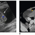
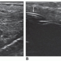
4,8 The posterior rectus sheath ends at the arcuate line, located halfway between the umbilicus and the pubis symphysis. The inferior, or distal, quarter of the rectus abdominus muscle is covered posteriorly by the transversalis fascia, which is all that separates the rectus muscles from the parietal peritoneum in the pelvic region10 (Fig. 4-7A, B).


The linea alba (or white line) is a midline, dense connective tissue structure that separates the right and left bellies of the rectus abdominus muscle. It is a fusion of the aponeuroses that form the rectus sheath.4,10

The linea alba extends from the xyphoid process to the pubic symphysis. Superiorly, the linea alba is wider and it narrows inferior to the umbilicus to the width of the pubic symphysis. The linea alba transmits small vessels and nerves to the skin (Figs. 4-4, 4-6A, and 4-7A, B). In thin, muscular people, a groove is visible in the skin overlying the linea alba.

The umbilicus is the area where all layers of the anterolateral abdominal wall fuse.4 The umbilical ring is a defect in the linea alba located deep to the umbilicus.4 This is the area through which the fetal umbilical vessels passed into the umbilical cord to connect with the placenta. The umbilicus and umbilical ring are the remnants of that fetal connection.

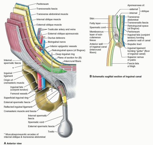
The inferior border of the external oblique aponeurosis extends between the anterior superior iliac spine and the pubic tubercle forming the inguinal ligament.10 Located in the inguinal region superior and parallel to the medial half of the inguinal ligament is the inguinal canal, a passageway through the abdominal wall that is formed during fetal development. It is an important canal where structures exit and enter the abdominal cavity, and the exit and entry pathways
are potential sites of herniation.4,10,11 In adults, the inguinal canal is an oblique passage approximately 4 to 6 cm long. Functionally and developmentally distinct structures located within the canal are the spermatic cord in males and the round uterine ligament in females. Other structures included in the canal in both sexes are blood and lymphatic vessels and the ilioinguinal and genital nerves.10 The entrance of the inguinal canal is formed by the deep inguinal ring at the superior end. The superficial (external) inguinal ring forms the exit at the inferior end. Normally, the inguinal canal is collapsed anteroposteriorly against the spermatic cord or round ligament. Between the two openings (rings), the inguinal canal has two walls (anterior and posterior), a roof, and a floor10,11 (Table 4-2 and Fig. 4-8A, B).


Posterior Abdominal Wall

The posterior abdominal wall is composed of the lumbar vertebra, posterior abdominal wall muscles, diaphragm, fascia, lumbar plexus, fat, nerves, blood vessels, and lymphatic vessels. On the posterior abdominal wall, the thoracolumbar fascia is an extensive complex. Medially, it attaches to the vertebral column. In the lumbar region, the thoracolumbar fascia has posterior, middle, and anterior layers with enclosed muscles between them. The fascia is thin and transparent in the thoracic region, whereas it is thick and strong in the lumbar region. The posterior (the most superficial) and middle layers of the thoracolumbar fascia enclose the bilateral erector spinae muscles or the vertical deep back muscles in the same manner that the rectus sheath encloses the rectus abdominus muscles.4 The thoracolumbar fascia is stronger than the rectus sheath because it is thicker and has a central attachment to the lumbar vertebrae. The rectus sheath has no bony attachment and fuses with the linea alba. The lumbar part of the posterior sheath, extending between the 12th rib and the iliac crest, attaches laterally to the internal oblique and transversus abdominis muscles. The thoracic portion attaches to the latissimus dorsi4 (see Fig. 4-9).

The anterior layer (which is the deepest layer) of the thoracolumbar fascia is the quadratus lumborum fascia, covering the anterior surface of the quadratus lumborum muscle.4 It is thinner than the middle and posterior layers of the thoracolumbar fascia. The anterior layer attaches to the anterior surfaces of the lumbar transverse processes, to the iliac crest, and to the 12th rib. Laterally, the anterior layer is continuous with the aponeurotic origin of the transversus abdominis muscle. Superiorly, it thickens to form the lateral arcuate ligament, and inferiorly, it is adherent to the iliolumbar ligaments4 (see Fig. 4-9).


Muscles

The muscles of the posterior abdomen are categorized as the superficial and intermediate extrinsic back muscles and the superficial layer, intermediate layer, and deep layer of intrinsic back muscles4 (Table 4-3). The three main, bilaterally paired, muscles comprising the posterior abdominal wall are the psoas major, iliacus, and quadratus lumborum (Fig. 4-10).


Layers

The posterior abdominal wall is covered with a continuous layer of endoabdominal fascia, which is continuous with the transversalis fascia.4 The posterior wall fascia is located between the parietal peritoneum and the muscles. The psoas fascia (sheath) is attached medially to the lumbar vertebrae and pelvic brim. Superiorly, the psoas fascia is thickened and forms the medial arcuate ligament. Laterally, the psoas fascia fuses with both the quadratus lumborum fascia and the thoracolumbar fascia. Inferior to the iliac crest, the psoas fascia is continuous with that part of the iliac fascia that covers the iliacus4 (see Fig. 4-9).


Diaphragm

The diaphragm is a fibromuscular domed structure separating the thoracic cavity from the abdominal cavity.4,12 The convex superior surface forms the floor of the thoracic cavity, and the concave inferior surface forms the roof of the abdominal cavity. The concave surfaces form the right and left domes with the right dome slightly higher because of the presence of the liver and the central part slightly depressed by the pericardium.4,12 The origin of the diaphragm is located at its periphery, which attaches to the inferior margin of the thoracic cage and the superior lumbar vertebrae.4,12 The diaphragm is the primary muscle inspiration. The central part descends during inspiration to enlarge the thoracic cavity and ascends during expiration to increase thoracic pressure. The diaphragm varies in postural position (supine or standing) and varies in height based on the size and degree of abdominal visceral distention.4,12

The muscular part of the diaphragm is located peripherally with fibers that converge radially on the central tendon. The central tendon has no bony attachments and appears incompletely divided into what resembles the three leaves of a wide cloverleaf. Although it lies near the center of the diaphragm, the central tendon is closer to the anterior part of the thorax4,12 (Fig. 4-11).

The area around the caval opening in the central tendon of the diaphragm is surrounded by a muscular part that forms a continuous sheet. The continuous sheet is divided into three parts based on its area of attachment: the sternal part, the costal part, and the lumbar part4,12 (Table 4-4).

The diaphragmatic crura are musculotendinous bands that arise from the anterior surfaces of the bodies of the superior three lumbar vertebrae, the anterior longitudinal ligament, and the intervertebral disks. The right crus is larger and longer than the left crus and appears as a triangular mass anterior to the aorta.4 It arises from the first three lumbar vertebrae and appears posterior to the caudate lobe of the liver. Fibers from the right crus extend anteriorly to form the esophageal hiatus.4,12 The left crus arises from the first two lumbar vertebrae.4


Diaphragmatic Apertures

The diaphragmatic apertures (openings, hiatus) permit several structures (esophagus, blood vessels, nerves, and lymphatic vessels) to pass between the thorax and the abdomen.4,12 The three larger apertures are the caval, esophageal, and aortic, and there are a number of small openings.12,13 The caval hiatus is primarily for the inferior vena cava (IVC) as it ascends into the thoracic cavity.4,13 The IVC shares the caval opening with the terminal branches of the right phrenic nerve and a few lymphatic vessels passing from the liver to the middle phrenic and mediastinal lymph nodes.4,13 Located to the right of midline, at the junction of the right and middle leaves of the central tendon, the caval opening is the most superior and anterior of the three large diaphragmatic apertures. Because the IVC is adherent into the margin of the caval opening, diaphragmatic contraction during inspiration widens the opening, which allows the IVC to dilate and helps facilitate blood flow through this large vein to the heart.4,12,13

The esophageal hiatus is an oval opening located in the muscle of the right crus anterior and superior to the aortic
hiatus.4,12 In 70% of individuals, both margins of the hiatus are formed by muscular bundles of the right crus. In 30% of individuals, a superficial muscular bundle from the left crus contributes to the formation of the right margin of the hiatus. The hiatus allows the esophagus to course from the thorax into the abdominal cavity and also serves as the passageway for the right and left trunks of the vagus nerve, esophageal branches of the left gastric vessels, and a few lymphatic vessels.4,12,13


The aortic hiatus passes between the crura posterior to the median arcuate ligaments at the inferior border of the T12 vertebra.4,12 This opening in the posterior diaphragm allows the descending aorta to course from the thoracic cavity to the abdominal cavity. The thoracic duct; azygos vein; and, sometimes, hemiazygos veins are also transmitted through the aortic hiatus. The aorta does not pierce the diaphragm or adhere to the hiatus, which means diaphragmatic movements during respiration do not affect aortic blood flow.4,12 The sternocostal triangle (foramen of Morgagni) is a small opening in the loose connective tissue between the sternal and costal attachments of the diaphragm. This triangle transmits lymphatic vessels from the hepatic diaphragmatic surface and the superior epigastric branch of the internal thoracic artery. The sympathetic trunk passes deep to the medial arcuate ligament and is accompanied by the least splanchnic nerves. In each crus, there are two small apertures: The right lesser aperture transmits the right greater and lesser splanchnic nerves and the left lesser aperture transmits the hemizygous vein (usually) and the left greater and lesser splanchnic nerves.12


Variants

Anatomic variants are composed of individual variations in fat and muscle content. In more muscular individuals, each lateral muscle layer tends to be identifiable, whereas in less well-developed individuals, muscle groups tend to be indistinct. Furthermore, some muscular components, like the psoas minor and pyramidalis muscles, are partially or completely absent in a significant portion of the population. It is important to note that in obese patients, the fatty layer variation can be significant.


SONOGRAPHIC APPEARANCES AND TECHNIQUES


Abdominal Wall

Sonography should be the first modality of choice for imaging abdominal wall structures because it is fast; widely available; and provides a valuable, inexpensive, and noninvasive method of imaging.7,14,15 The ability to perform real-time imaging that demonstrates changes in anatomy with changes in patient position and other maneuvers makes sonography ideal for evaluating structures in motion. Imaging the normal abdominal wall and detecting pathologic processes such as inflammatory lesions, hemorrhage, hernia, or masses makes sonography of the abdominal wall an excellent modality for diagnosing pathology. Many clinical questions can be answered with the use of sonography in evaluating posttrauma or postsurgical patients. It is extremely important to understand the normal sonographic appearance of the abdominal wall and the appropriate instrumentation and scanning techniques necessary to achieve that appearance.

Only gold members can continue reading. Log In or Register to continue

Stay updated, free articles. Join our Telegram channel

Dec 10, 2022 | Posted by in ULTRASONOGRAPHY | Comments Off on The Abdominal Wall and Diaphragm

Full access? Get Clinical Tree

Get Clinical Tree app for offline access