Fig. 13.1
Classical Seldinger technique for radial puncture. (a) Local anesthesia, (b) puncture of the radial with IV catheter, (c) blood back in mandrin, (d) 0.035″ guidewire inserted into IV sheath, (e) regular 5F sheath inserted, (f) vasodilator cocktail
Introducing and Exchanging Coronary Catheters
Insertion of the catheter is performed over a 150-cm J-tip fixed core wire. Fluoroscopy is generally advised as there is a risk that the J-tip inadvertently deflects into the common carotid or internal mammary arteries. Further, having the patient perform a breath hold in deep inspiration will aid the entry of the J-tip wire into the ascending aorta. Another maneuver that will assist the passage of the J-tip wire to the coronary cusps is by clocking the catheter (from RRA), which will turn the catheter away from the descending aorta.
Catheter exchanges can be performed over the 150-cm J-tip wire without the need for exchange-length wires. Having the J-tip wire resting in the coronary cusp, withdrawal of the catheter over the wire is performed until the distal end of the wire just disappears into the hub of the catheter. Next, a 10- or 20-mL Luer lock syringe filled with normal saline is attached followed by simultaneous injection of saline and withdrawal of the catheter, leaving the J-tip wire in place. The maneuver can be performed under fluoroscopy, but experienced operators usually do this without monitoring the tip of the wire. Occurrence of premature systolic contractions will indicate passage of the wire though the aortic valve, which will be corrected by retracting the wire under fluoroscopy. The next catheter is introduced over the 150-cm wire while ensuring its stable position in the coronary cusp. In patients with dilated ascending or unfolded aortas, there is a risk that the proximal third of the J-tip wire prolapse towards the descending aorta, resulting in further advancement of the wire, without externalization from the proximal hub of the catheter. Although such situations could lead to an embarrassing situation of “losing” a wire into the patient, a simple withdrawal of the catheter outside the introducer will invariably expose the proximal end of the J-tip wire. The catheter can then be reinserted on the wire, with an alternative technique of proximal wire control. However, occasional fluoroscopy to ensure the J-tip position will help overcome this difficulty.
Inability to Cannulate the Wire or to Advance the First Catheter
First and foremost is to confirm an arterial location of the sheath by connecting the side arm to the arterial monitoring system. Failure to establish pulsatile flow can indicate either distal spasm at the tip of the introducer, radial artery dissection, false lumen catheter insertion, or venous insertion. If the pulsatile flow was established, but the wire cannot progress beyond the forearm, the operator is likely facing a radial loop or a spasm. Alternatively, wire passage into the brachioradial artery (radial artery with a high origin) or the recurrent radial artery [8] can explain why the catheter will not progress, but this is an unusual situation. As loops and spasms explain most of the difficulties, we tend to simply probe the radial with a Bentson guidewire to see if it will successfully traverse the radial to the brachial artery. Alternatively, a Glydewire can be used, keeping in mind that this type of wire is more prone to engage radial artery side braches and cause perforations. The authors prefer the use of a coronary workhorse guidewire, with an atraumatic coil tip, to navigate through loops and spastic segments, usually with success. Micro-perforations commonly occur after extensive wire manipulation, followed by the performance of a forceful angiography to document the etiology of difficult wire cannulation resulting in a hydraulic worsening of any small wire tip perforation. We thus reserve forearm angiography only to situations where all the earlier steps have failed. If an angiography is warranted, a very careful low-pressure injection of contrast should be performed. Vasospasm can also be managed with a further dose of antispasmolytic agents and/or further sedation, especially if noticed on angiography. Changing the catheter to one of a smaller caliber or to one with a gentler curve (e.g., JR, MP) can assist in negotiating tortuosity. Most importantly, the operator should never force the wire or the catheter. On a rare occasion, the operator has to abandon the radial approach and convert to a contralateral access or the femoral approach. During the procedure in the absence of forearm swelling, should a small perforation of the radial or brachial artery be detected, we generally advise to continue as the presence of the catheter helps seal the perforation. Other techniques include inserting a long arterial sheath [9] or sealing it with a prolonged inflation of a peripheral arterial balloon [10].
Choice of Diagnostic Catheters
Related posts:
 Adjunctive Pharmacology for Coronary Intervention
 Percutaneous Coronary Interventions in the Elderly
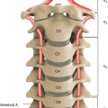 Percutaneous Management of Carotid and Vertebral Artery Disease
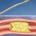
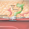 Vascular Access, Closure, and Management
 Percutaneous Cell Therapy for Acute and Chronic Cardiac Disease
 Application of Advanced Technologies for the Treatment of Lower Extremity Arterial Disease
Stay updated, free articles. Join our Telegram channel

Full access? Get Clinical Tree


