7 The upper limb
The bones of the upper limb
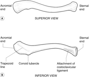
The scapula (Figs 7.1, 7.2)
This flat triangular bone has three processes:
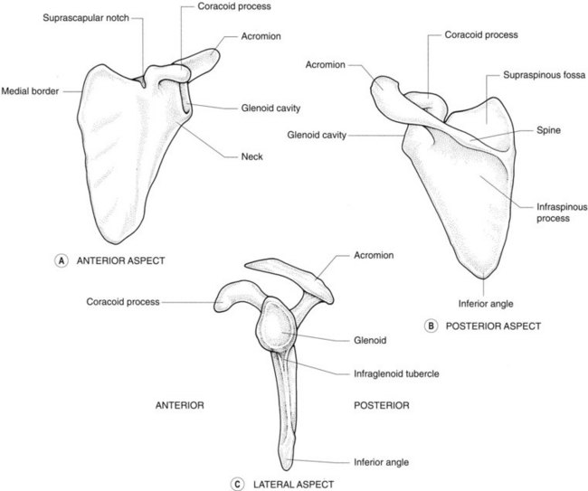
Radiological features of the scapula
Plain radiographs
The inferior angle of the scapula lies over the seventh rib or interspace – this is a useful guideline in identifying ribs or thoracic vertebral levels.
The scapula lies over the ribs and obscures some of the lung fields in PA chest radiographs unless the shoulders are rotated forwards. In AP views it is not usually possible to rotate the scapulae off the lung fields. Similarly, in AP views of the scapula the beam is centred over the head of the humerus to project the thoracic cage away from the scapula.
In lateral chest radiographs, the lateral border of the scapula may be confused with an oblique fissure. The inferior angle of the scapula may be slightly bulbous and simulate a mass on this view.
Isotope bone scan
The inferior angle of the scapula overlying the seventh rib may appear as a ‘hot spot’.
Ossification
The scapula ossifies in the eighth week of fetal life. An ossification centre appears in the middle of the coracoid process in the first year of life and fuses at 15 years of age. Secondary centres appear in the root of the coracoid process, the medial border and the inferior angle of the scapula between 14 and 20 years, and fuse between 22 and 25 years of age.
Radiology pearl
The glenoid is oblique from posterior to anterior, lateral to medial in orientation. This is an anatomic evolutionary feature that prevents posterior dislocation following fall on an outstretched hand. The same feature leads to the tendency to dislocate anteriorly following a direct fall on the shoulder.
Radiological features of the clavicle
Chest radiograph
The clavicle overlies the apices of the lungs in chest radiographs. Apical or lordotic views are used to project the clavicles above the lungs to evaluate this area further. In portable AP chest radiography, if the patient is inclined backwards from a true vertical position the horizontal beam projects the clavicles above the lungs.
On a chest radiograph, the distance between the medial end of the clavicle and the spine of the vertebrae is equal on both sides unless the patient is rotated.
Ossification
The clavicle begins to ossify before any other bone in the body. It ossifies in membrane from two centres that appear at the fifth and sixth fetal weeks, and fuses in the seventh week. A secondary centre appears at the sternal end at 15 years in females and 17 years in males, and fuses at 25 years of age.
Radiology pearl
Being membranous rather than arising from a chondral frame (endochondral), cartilage tumours of the clavicle are extremely uncommon.
Multislice CT with reformatted images allows excellent tomographic assessment of the long axis of the clavicle. The articulation with the sternum is best visualized using MRI with a surface coil placed over the anterior chest wall.
The humerus (Figs 7.4, 7.5; see also Fig. 7.2)
The shaft is marked by a spiral groove where the radial nerve and the profunda vessels run. The deltoid tuberosity on the lateral aspect of the midshaft is the site of insertion of the deltoid muscle.
The lower end of the humerus is expanded and has medial and lateral epicondyles. The articular surface for the elbow joint has a capitellum for articulation with the radial head and a trochlea for the olecranon fossa of the ulna. Above the trochlea are fossae, the coronoid anteriorly and the deeper olecranon fossa posteriorly.
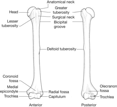
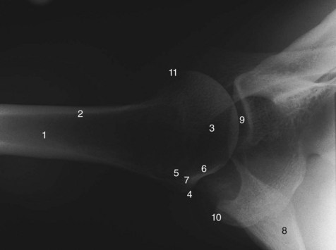
Radiological features of the humerus
Plain radiographs
The lower epiphysis of the humerus lies at a 25° angle to the shaft so that a vertical line down the front of the shaft on a lateral radiograph – the anterior humeral line – bisects the capitellum.
An olecranon foramen may replace the olecranon fossa.
Radiology pearl
A hook-shaped projection of bone – termed the supracondylar process – occasionally occurs about 5 cm above the medial epicondyle. It varies in length between 2 and 20 mm and may be continuous with a fibrous band, the ligament of Struthers, attached above the epicondyle to form a foramen that transmits the median nerve and the brachial artery.
Avulsion of the medial epicondyle
The flexor muscles of the forearm arise from the medial epicondyle of the humerus. Repeated contractions or a single violent contraction of these muscles in a child can result in avulsion of the apophysis (a secondary ossification centre occurring outside a joint) of the medial epicondyle.
Ossification
The primary centre for the humerus appears at the eighth week of fetal life. Secondary centres appear in the head of the humerus at 1 year, the greater tuberosity at 3 years, and the lesser tuberosity at 5 years of age. These fuse with one another at 6 years and with the shaft at 20 years of age. Secondary centres appear in the capitellum at 1 year, the radial head at 5 years, the internal epicondyle at 5 years, trochlea at 10 years, olecranon at 10 years and external epicondyle at 10 years (CRITOE). These fuse at 17–18 years of age.
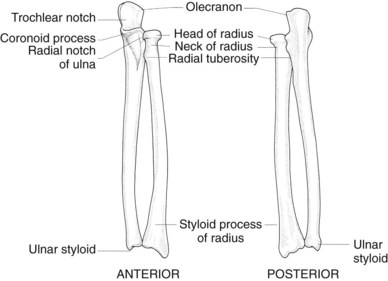
The radius and ulna (Figs 7.6, 7.7; see also Fig. 7.5)
The upper part of the ulna – the olecranon – is hook-shaped, with the concavity of the hook – the trochlear fossa – anteriorly. A fossa found laterally at the base of the olecranon is for articulation with the radial head. The shaft of the ulna is narrow. The styloid process at the distal end is narrower and more proximal than that of the radius.
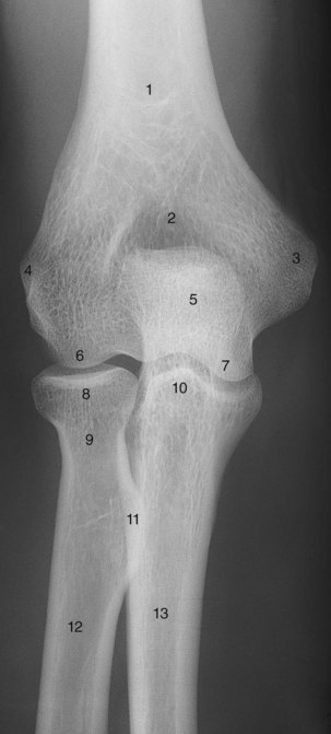
Radiological features of the radius and ulna
Plain radiographs
The head of the radius has a single cortical line on its upper surface and is perpendicular to the neck in the normal radiograph (see Fig. 7.5). Angulation of the head or a double cortical line are signs of fracture of the radial head.
The ulnar styloid is proximal to the radial styloid, with a line joining them on an AP radiograph lying at an angle of 110° with the long axis of the radius (see Fig. 7.7). In a lateral radiograph, the articulating surface of the distal radius is angled 10° to a line through the shaft of the radius. Recognition of these normal angles is important in reduction of fractures of the wrist.
Radiology pearl
Failure to restore volar angulation of the distal radius following a fracture of the distal radius often results in loss of grip strength due to impaired flexor function.
The pronator quadratus is a square, flat muscle that arises on the distal ulna and passes to the distal radius. A thin fat pad overlying this muscle is visible as a linear lucency on a lateral radiograph of the wrist. Thickening of the muscle, such as by haematoma in fracture of the underlying bone, can be detected on a radiograph by bowing of the pronator quadratus fat pad.
Ossification of the radius
The primary ossification centre of the radius appears in the eighth week of fetal life. Secondary centres appear distally in the first year and proximally at 5 years of age. These fuse at 20 years and 17 years, respectively.
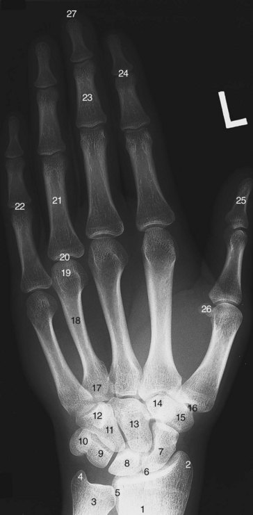
The carpal bones (Fig. 7.8; see also Fig. 7.7)
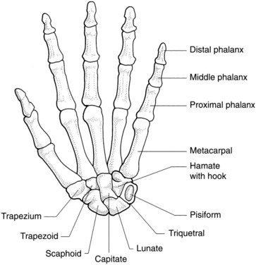
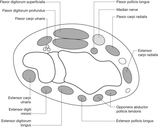
Together the carpal bones form an arch, with its concavity situated anteriorly. The flexor retinaculum is attached laterally to the scaphoid and the ridge of the trapezium, and medially to the pisiform and the hook of the hamate. It converts the arch of bones into a tunnel, the carpal tunnel, which conveys the superficial and deep flexor tendons of the fingers and the thumb (except flexor carpi ulnaris and palmaris longus tendons) and the median nerve. The extensor retinaculum on the dorsum of the wrist attaches to the pisiform and triquetrum medially and the radius laterally. Six separate synovial sheaths run beneath it (Fig. 7.9).
Radiological features of the carpal bones
Radiography
These are radiographed in the anteroposterior, lateral and oblique positions (see Fig. 7.7). Carpal tunnel views are obtained by extending the wrist and taking an inferosuperior view that is centred over the anterior part of the wrist.
Supernumerary bones
These may be found in the wrist and include the os centrale found between the scaphoid, trapezoid and capitate, which may represent the tubercle of the scaphoid that has not fused with its upper pole, and the os radiale externum, which is found on the lateral side of the scaphoid distal to the radial styloid.
Nutrient arteries of the scaphoid
In 13% of subjects these enter the scaphoid exclusively in its distal half. If such a bone fractures across its midportion, the blood supply to the proximal portion is cut off and ischaemic necrosis is inevitable. This occurs in 50% of patients with displaced scaphoid fractures.
Ossification of the carpal bones
These ossify from a single centre each. The capitate ossifies first and the pisiform last, but the order and timing of the ossification of the other bones is variable. Excluding the pisiform, they ossify in a clockwise direction from capitate to trapezoid as follows: the capitate at 4 months; the hamate at 4 months; the triquetral at 3 years; the lunate bone at 5 years; and the scaphoid, trapezium and trapezoid at 6 years. The pisiform ossifies at 11 years of age.
The metacarpals and phalanges
The five metacarpals are numbered from the lateral to the medial side. Each has a base proximally that articulates with that of the other metacarpals, except in the case of the first metacarpal, which is as a result more mobile and less likely to fracture. The third metacarpal has a styloid process extending from its base on the dorsal aspect. Each metacarpal has a rounded head distally, which articulates with the proximal phalanx.
The phalanges are 14 in number, three for each finger and two for the thumb. Like the metacarpals, each has a head, a shaft and a base. The distal part of the distal phalanx is expanded as the tuft of the distal phalanx.
Radiological features of the metacarpals and phalanges
Bone age
A radiograph of the left hand is used in the determination of bone age. Standards of age determined by epiphyseal appearance and fusion have been compiled for the left hand and wrist by Greulich and Pyle, and by Tanner and Whitehouse (TW2 method).
The metacarpal sign
A line tangential to the heads of the fourth and fifth metacarpals does not cross the head of the third metacarpal in 90% of normal hands – this is called the metacarpal sign. This line does, however, cross the third metacarpal head in gonadal dysgenesis.
The carpal angle
This is formed by lines tangential to the proximal ends of the scaphoid and lunate bones. In normal hands the average angle is 138°. It is reduced to an average 108° in gonadal dysgenesis.
The metacarpal index
This is calculated by measuring the lengths of the second, third, fourth and fifth metacarpals and dividing by their breadths taken at their exact midpoint. The sum of these divided by four is the metacarpal index, which has a normal range of 5.4–7.9. An index greater than 8.4 suggests the diagnosis of arachnodactyly.
Sesamoid bones
Two sesamoid bones are found related to the anterior surface of the metacarpophalangeal joint of the thumb in the normal radiograph. A single sesamoid bone in relation to this joint in the little finger is seen in 83% of radiographs, and at the interphalangeal joint of the thumb in 73%. These are occasionally found at other metacarpal and distal interphalangeal joints. The incidence of sesamoid bones is increased in acromegaly.
Ossification of the metacarpals and phalanges
These ossify between the ninth and twelfth fetal weeks. Secondary ossification centres appear in the distal end of the metacarpals of the fingers at 2 years and fuse at 20 years of age. Secondary centres for the thumb metacarpal and for the phalanges are at their proximal end and appear between 2 and 3 years, and fuse between 18 and 20 years of age.
The joints of the upper limb
The sternoclavicular joint
Articular surfaces
These are the sternal end of the clavicle, the clavicular notch of the manubrium and the upper surface of the first costal cartilage.
The acromioclavicular joint
Articular surfaces
These are the outer end of the clavicle and the acromion. In health the undersurface of the acromion will align with the undersurface of the clavicle.
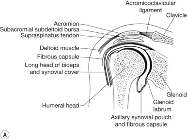
The shoulder (glenohumeral) joint (Fig. 7.11)
Articular surfaces
The articular surface of the humeral head is four times the area of the glenoid cavity.
Capsule
This is attached to the epiphyseal line of glenoid and humerus, except inferiorly where it extends downwards on the medial aspect of the neck of the humerus as the axillary pouch.
Synovium
In addition to lining the capsule of the joint, the synovium extends along the tendon of the long head of the biceps and beneath the tendon of subscapularis muscle as the subscapular bursa. The long head of the biceps is therefore extrasynovial but intracapsular attaching to the supraglenoid tubercle.
Ligaments
Stability
In addition to ligaments, the stability of the shoulder joint depends upon the surrounding muscles. These are:
The inferior part of the joint is least well protected by either ligaments or muscles.
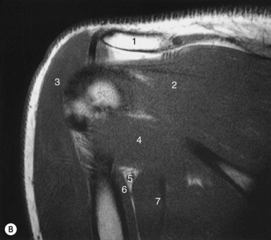
Radiological features of the shoulder joint
Plain radiographs
The supraspinatus muscle passes on the superior aspect of the shoulder joint to the greater tuberosity of the humerus. Calcification occurs in this muscle owing to degenerative change and may be visible on radiographs.
The supraspinatus muscle is separated from the acromion by the subacromial–subdeltoid bursa, the largest bursa in the body. This bursa does not communicate with the shoulder joint unless supraspinatus is ruptured by trauma or degeneration. This communication is then visible on arthrography. MRI can also be used to detect this rupture.
The capsule of the shoulder joint is lax and relatively unprotected by ligaments or muscles inferiorly. This is the site of accumulation of fluid in effusion or haematoma of the joint.
Arthrography with or without CT
In the shoulder joint, arthrography is achieved by injection of contrast into the joint below and lateral to the coracoid process. It shows the features of the joint as outlined above. In particular, the axillary pouch can be seen inferior to the humeral head on external rotation of the arm, and the subscapular (subcoracoid) bursa can be seen on internal rotation of the arm. The subacromial (subdeltoid) bursa is not filled unless the supraspinatus tendon is completely ruptured.
The tendon of the long head of biceps is seen as a filling defect within the joint and its synovial sheath is opacified outside the joint along the bicipital groove of the humerus.
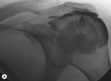
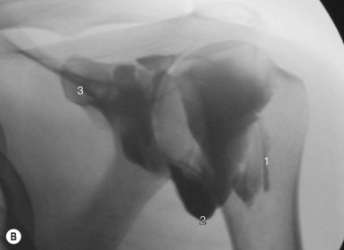
Subacromial bursography
Contrast injection to the subacromial bursa with either ultrasound or fluoroscopic guidance is often followed by therapeutic injection of steroid and bupivacaine (Marcain). Subacromial bursitis or inflammation of the bursa is most frequently secondary to impingement (Fig. 7.10A, B).
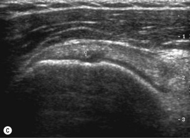


Figure 7.10 • (A) Contrast outlining the subacromial bursa allowing targeted placement of therapeutic steroid. (B) Shoulder arthrogram showing contrast filling of joint space. (C) High-frequency ultrasound showing supraspinatus tendon attachment to humeral head. (D) Corresponding coronal oblique MRI.
Ultrasound
High-frequency linear probes are now often employed as an alternative to MRI to evaluate the rotator cuff (Fig. 7.10C, D).
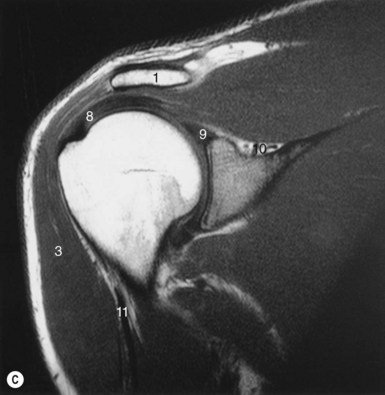
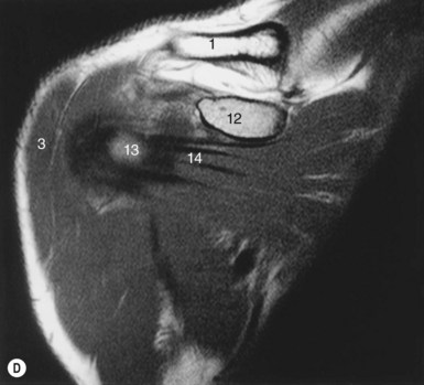
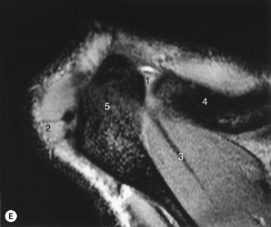
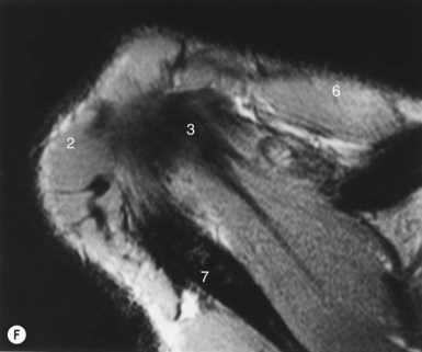
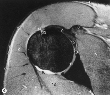
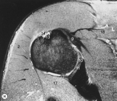
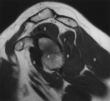
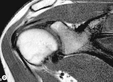
Magnetic resonance imaging (Figs 7.11, 7.12)
MRI with surface coils is used increasingly to image the shoulder joint.


Figure 7.11 • (A) Coronal oblique section through the shoulder joint as seen on coronal oblique MRI scan. (B, C, D) Coronal oblique T1-weighted images from posterior to anterior.
(E, F, G, H) Direct axial images of the shoulder from superior to inferior.

Full access? Get Clinical Tree

