Age/gender
Histology/location
Treatment before/after targeted RT*
Total activity (MBq)/radionuclide
Barthel index before/at the end of targeted RT
Survival after initiation
of targeted RT
Overall sur-vivala
Glioblastomas
37/F
G/fL
S-R-C
375/213Bi
80/70
7
26
52/M
G/ftR
S-C/−
5250/90Y
100/95
24
25
60/F
G/pL
R-S/S
5250/90Y
95/95
18
23
21/F
G/pons
*R-C/−
1125/177Lu
15/70
10
20
66/F
G/pL
S/C
3750/90Y
100/90
18
19
57/F
G/poL
*S/−
5625/90Y
95/90
18
19
47/F
G/für
S-R-C/S
4500/90Y
5/40
9
17
69/M
G/tR
S/−
4688/90Y
90/90
15
16
50/M
G/bifcR
S-C-R
1875/90Y
25/35
8
15
40/M
G/fR
S/−
7500/90Y
100/95
13
14
63/M
G/ftR
S-R-C/−
5250/90Y
30/50
11
14
44/F
G/thL
*−/−
4875/90Y
15/65
8
9
71/F
G/tpL
*−/−
3750/90Y
100/65
6
7
70/M
G/fL
S/−
7500/90Y
100/75
6
7
Gliomas WHO grades 2–3
37/F
OAIII/thR
S-C/−
2250/177Lu
40/75
+20
+149
42/M
OII/pcL
S/S
825/213Bi
90/90
+66
+67
34/M
AII/pfL
S/S
7500/90Y
100/100
+26
+41
63/M
AIII/toL
*R-C/S
6375/177Lu
60/95
11
28
34/M
OII/fR
*−/S
5625/90Y
100/100
+28
+29
31/M
OII/fL
*−/S
2250/90Y
100/100
+22
+23
The application of the radiopharmaceutical was straightforward. The radiopharmeutical was distributed according to tumor geometry. Only transient toxicity was seen as symptomatic radiogenic edema in one patient. The observation period ranged from 7 to 66 months after completion of targeted radiopeptide therapy. Disease stabilization and/or improved neurologic status was observed in 13 of 20 patients. Secondary resection disclosed widespread radiation necrosis with improved demarcation of the tumor.
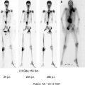
In addition to the preclinical and clinical evaluation of DOTATAGA-SP, a more sophisticated dosimetry protocol was applied to this novel therapy option (Kneifel et al. 2007). The aim was to establish a reproducible dosimetry for intratumoral radiopeptide therapy to be able to compare the doses of this modality to external beam radiotherapy and to identify the effective dose range. In 12 patients with malignant gliomas 2 MBq of 111In-substance P and 370–3,330 MBq of 90Y-substance P were applied subsequently, and each time serial SPECT scans were acquired over a period of 24 h. Quantitative voxelwise dose distribution maps (in Gy/GBq) were computed from these data. The correlation between pre- and post-therapeutic dosimetry was accurate. The calculated absorbed dose to the tumor was found to be as high as 40–483 Gy/GBq (mean 155 Gy/GBq) for the first therapeutic injection with 90Y-substance P. Given an average therapeutic activity of 30 mCi (1.11 GBq) this results in a mean absorbed radiation dose of 172 Gy within the tumor.
Based on these encouraging results two further studies were initiated with radiolabeled SP.
First of all it was analyzed whether neoadjuvant treatment prior to tumor resection is also a valid concept to improve therapy of malignant brain tumors (Cordier et al. 2010). The previous study (Kneifel et al. 2006) showed, that from a surgical point of view, resectability was facilitated due to improved demarcation and the radiation-induced anti-angiogenic effect in these patients who had been treated upfront with loco-regional radiopeptide therapy.
A total of 17 glioblastoma (GBM) patients were treated by local injection of 90Y-DOTAGA-substance P prior to tumor resection. Patients were treated by four cycles of intracavitary radiopeptide therapy with 90Y-DOTAGA-substance P in monthly intervals. In case of suspected tumor recurrence on imaging studies, confirmation or exclusion by biopsy was offered. The cumulative injected activity ranged from 100 to 345 mCi. A summary of the therapy scheme as well as the results of this study is given in Table 2.
Table 2
.
Age/gender | GBM location | Cycles/cumulative dose (mCi) | Post-OP adjuvant therapy | Barthel index $ (pre/post) | Pro-gression free survival (months) | Overall survival (months) |
|---|---|---|---|---|---|---|
50/F | po,R
Related posts: (Yttrium-90 Microspheres) for Metastatic Hepatic Malignancies (Yttrium-90 Microspheres) for Metastatic Hepatic Malignancies
 Thyroid Carcinoma Thyroid Carcinoma
 of Progressive Dedifferentiated and Medullary Thyroid Cancer with Radiolabeled Somatostatin Analogs of Progressive Dedifferentiated and Medullary Thyroid Cancer with Radiolabeled Somatostatin Analogs
 Bisphosphonates for Bone Pain Palliation Bisphosphonates for Bone Pain Palliation
 Radiopharmaceuticals for PET Dosimetry Before, During, and After Endoradiotherapy Radiopharmaceuticals for PET Dosimetry Before, During, and After Endoradiotherapy
 Use of Dosimetry in the Planning of Patient Therapy Use of Dosimetry in the Planning of Patient Therapy
Stay updated, free articles. Join our Telegram channel
Full access? Get Clinical Tree
 Get Clinical Tree app for offline access

|