Fig. 1
In 1995, a 58-year-old gentleman received combined chemoradiation for small cell lung cancer of the right lung (limited disease) resulting in a cure. (a) Shows a follow-up computed tomography (CT) scan from 2008 demonstrating slight paramediastinal radiation fibrosis. In January 2009, the patient was diagnosed with stage IIIB adenocarcinoma of the right lung (b). He received two cycles of platinum-based chemotherapy and achieved a partial response. He went on to simultaneous chemotherapy and 3D-conformal reirradiation (15 fractions of 2.8 Gy). He developed grade III acute esophagitis but no pulmonary toxicity. (c) Shows the most recent CT scan taken 1 year after reirradiation demonstrating increasing radiation fibrosis in the reirradiated region (clinically asymptomatic). At that time the patient was diagnosed with multiple brain metastases and started palliative whole-brain irradiation (no previous prophylactic whole-brain radiotherapy had been given)
3.5 Spinal Cord
Reirradiation tolerance of the spinal cord has been most extensively studied in animal models using paresis, predominantly resulting from white matter necrosis that manifests in 10–12 months in rodents, as the end point and the median paresis dose (ED50) for computation. Results of two reirradiation experiments revealed that the fractionation sensitivity of reirradiation was similar to that of single-course irradiation (Ruifrok et al. 1992a; Wong et al. 1993). Lower spine reirradiation experiments (T10-L2 in adult mice, L3–5 in adult rats, L2–6 in young guinea pigs) showed remarkable long-term recovery if the initial dose was limited to 50–75 % of the ED50 (Hornsey et al. 1982; Lavey et al. 1994; Knowles 1983; Mason et al. 1993). The extent of injury retained was highest after initial irradiation to 75 % of the ED50, but even under this condition, only 30 % of injury was retained. A series of reirradiation experiments of rat cervical spinal cord showed that the initial dose, the interval to reirradiation and the reirradiation dose influenced the latency to myelopathy (Wong et al. 1993; Wong and Han 1997). Beyond an 8-week interval, there was a progressive increase in recovery with an increasing time interval to reirradiation, but recovery was never complete. In adult rats main long-term recovery occurred between 2 and 6 months. For 3-week-old rats, partial recovery took place during the first month, which increased only slightly between 1 and 6 months (Ruifrok et al. 1992b). In a different study, an initial single dose of 15 Gy was administered, followed 8 or 16 weeks later by additional graded doses (van der Kogel 1979). In contrast to all other studies, separate dose-response curves were obtained for white matter necrosis (latent period <7 months) and vascular damage (latent period up to 18 months). Recovery between 8 and 16 weeks was significant for the white matter necrosis end point but was much less for vascular damage. It has also been shown that modification of the reirradiation tolerance of the spinal cord can be obtained (Nieder et al. 2005a). In these experiments, a combination of systemically administered insulin-like growth factor-1 and intrathecal amifostine resulted in lower incidence of myelopathy after cervical spinal cord reirradiation in rats. Such strategies were not pursued further because of increasing availability of equipment and software, which allows for spinal cord sparing (IMRT, stereotactic RT etc.). Other pharmacologic strategies aiming at radioprotection in different tissues and organs typically were examined in previously untreated animals (Greenberger 2009) although the reirradiation setting appears attractive for studying radioprotectors.
As compared to rodent experiments, more clinically applicable data were generated in adult rhesus monkeys at MD Anderson Cancer Center, Houston, USA, by Ang et al. (1995). The cervical spinal cord of these primates was irradiated with 2.2 Gy per fraction. The control group received a single radiation course to total doses of 70.4, 77 or 83.6 Gy. The number of animals developing myelopathy in these 3 dose groups was 3 of 15, 3 of 6 and 7 of 8, respectively. Twelve asymptomatic animals in the 70.4 Gy group were retreated 2 years later to cumulative doses of 83.6, 92.4 or 101.2 Gy (four animals each). Only two developed paresis at 11 (83.6 Gy) and 8 (92.4 Gy) months after reirradiation. Subsequently, 16 monkeys received 44 Gy in 20 fractions and 2 years later were reirradiated to cumulative doses of 83.6, 92.4, 101.2 or 110 Gy. Only two (one received 101.2 Gy and the other 110 Gy) developed myelopathy. These data indicate that a substantial amount of the occult injury induced by the priming dose decayed within 2 years. As published in 2001, Ang et al. confirmed their initial observations in a group of 56 rhesus monkeys. The dose of the initial course was 44 Gy in all monkeys. Reirradiation dose was 57.2 Gy, given after 1-year (n = 16) or 2-year (n = 20) intervals, or 66 Gy, given after 2-year (n = 4) or 3-year (n = 14) intervals. Only 4 of 45 monkeys completing the required observation period (2–2.5 years after reirradiation, 3–5.5 years total) developed myelopathy. Fitting the data with a model, assuming that all (single course and reirradiation) dose-response curves were parallel, yielded recovery estimates of 33.6 Gy (76 %), 37.6 Gy (85 %) and 44.6 Gy (101 %) of the initial dose, after 1, 2 and 3 years, respectively, at the 5 % incidence level. Another way to look at these results is to estimate the total cumulative dose that can be tolerated, expressed in EQD2 that is equivalent dose in 2-Gy fractions calculated using the linear-quadratic approach. For a time interval of 1, 2 and 3 years between the treatment courses, cumulative doses of 150, 156 and 167 % of the first-line setting’s tolerance dose appear possible.
If true in humans, an initial exposure equivalent to 46 Gy in 2-Gy fractions (arbitrarily selected to represent 100 % of the tolerance dose at the 5 % myelopathy risk level because many institutions limit the spinal cord dose to lower levels than true tolerance (Kirkpatrick et al. 2010)) might be followed by an additional 23–24 Gy in 2-Gy fractions (50 % of the tolerance dose) 1 or 2 years later. Clinical data from different institutions supporting this interpretation have been published (Schiff et al. 1995; Grosu et al. 2002). Most patients were treated with palliative reirradiation and therefore follow-up was often limited. In patients with better prognosis, the cumulative spinal cord dose often was kept at very low levels, e.g. in the RTOG head and neck cancer reirradiation protocols (Langer et al. 2007). Nevertheless, data from patients with longer follow-up have been published, e.g. five patients with recurrent Hodgkin’s disease who were followed for more than 5 years after reirradiation (Magrini et al. 1990). The first spinal cord dose was 30 Gy in 1.7 Gy fractions (plus chemotherapy) and 1–3 years later up to 40 Gy in 2-Gy fractions was administered (two to three vertebral segments) without causing myelopathy. The cumulative EQD2 of this treatment is slightly lower than 150 %. All available data from different published series including those reporting on myelopathy (Wong et al. 1994) were analysed by Nieder et al. (2005b, 2006a). Seventy-eight patients were included and a risk prediction model was developed, based on time interval, cumulative dose and presence or absence of any treatment course resulting in quite high spinal cord exposure. Besides cumulative dose, interval <6 months and total dose equivalent to >50 Gy in 2-Gy fractions in one of the two courses increases the risk of myelopathy. Low-risk patients had <5 % risk of myelopathy and intermediate risk patients approximately 25 %. However, unpublished cases treated after 2006 in the author’s institution in Bodø, Norway, all remained free from myelopathy, suggesting that the actual risk might be lower than previously anticipated. Chapter “Fractionation Concepts” of this book contains a table showing calculations of the myelopathy risk based on this model (Table 3).
The introduction of stereotactic body radiotherapy has resulted in new challenges as such treatment typically is administered with few high-dose fractions or even single doses. Rather than irradiating complete spinal cord cross sections with homogeneous doses, small areas are exposed and steep dose gradients achieved. Medin et al. (2012) developed a swine model where a 10-cm length of the spinal cord (C3-T1) was uniformly irradiated to 30 Gy in 10 fractions and reirradiated 1 year later with a single radiosurgery dose centred within the previously irradiated segment. Radiosurgery was delivered to a cylindrical volume approximately 5 cm in length and 2 cm in diameter, which was positioned laterally to the cervical spinal cord, resulting in a dose distribution with the 90 %, 50 % and 10 % isodose lines traversing the ipsilateral, central and contralateral spinal cord, respectively. Follow-up after reirradiation was 1 year. Summarised briefly, pigs receiving radiosurgery 1 year following 30 Gy in 10 fractions were not at significantly higher risk of developing motor deficits than pigs that received radiosurgery alone. Limited data on reirradiated patients are available. Damast et al. (2011) reviewed the records of patients with in-field recurrence after previous spine radiation (median dose 30 Gy) that received salvage IMRT with either five 4-Gy (20-Gy group, n = 42) or five 6-Gy (30-Gy group, n = 55) daily fractions. The median follow-up was 12 months (range, 0.2–63.6 months). Maximal point dose to the spinal cord and cauda equina were limited to 14 Gy and 16 Gy, respectively. Neither previous cord dose from first radiotherapy nor interval between first and second course had bearing on these dose constraints. There was no incidence of myelopathy. Notably, there were nine vertebral body fractures and one benign oesophageal stricture after IMRT, which was potentially attributable to radiation. In 36 patients treated with helical tomotherapy, radiation myelopathy was not observed (Sterzing et al. 2010). The most common initial regimen was 10 fractions of 3 Gy. All patients had low cumulative EQD2 and thus belonged to the low-risk group. Choi et al. (2010) retrospectively reviewed 51 lesions in 42 patients whose spinal metastases recurred in a previous radiation field (median previous spinal cord dose of 40 Gy) and were subsequently treated with stereotactic radiosurgery to a median marginal dose of 20 Gy (range, 10–30 Gy) in 1–5 fractions (median, 2). The median follow-up was 7 months (range, 2–47 months). The median spinal cord maximum total dose and dose per fraction from reirradiation were 19.3 Gy (range, 5.1–31.3 Gy) and 7.2 Gy (range, 2.9–19.3 Gy), respectively. One patient (2 %) experienced grade 4 neurotoxicity. Having received 39.6 Gy in 1.8 Gy fractions (total spinal cord dose of 40 Gy) to the T4 to L1 vertebral bodies 81 months before receiving reirradiation, she received 20 Gy in 2 fractions to treat a T5 recurrence. Treatment was prescribed to the 80 % isodose line. The maximal spinal cord dose from reirradiation was 19.25 Gy, which is equivalent to 56 Gy in 2-Gy fractions. Therefore, this patient would be predicted to have a high likelihood of myelopathy by the aforementioned risk score (Nieder et al. 2006a). A case with comparably high reirradiation dose (maximum 20.9 Gy in 2 fractions to the thecal sac at T1) resulting in myelopathy 5 months after retreatment has been reported by Sahgal et al. (2012). Previous treatment was only 25 Gy in 28 fractions (interval 70 months). A third case was also reported in the same paper. Here, initial treatment was close to tolerance, i.e. equivalent to 52 Gy in 2-Gy fractions. The maximum reirradiation dose to the thecal sac was 14.7 Gy in one single fraction (level T10, interval 12 months). Myelopathy developed after 3 months. In a fourth case, initial treatment was equivalent to 50 Gy in 2-Gy fractions and reirradiation dose maximum to the thecal sac was 32.6 Gy in 3 fractions (level C1/C2, interval 18 months). Myelopathy developed after 8 months. In their report, Sahgal et al. (2012) provided a recommendation of reasonable stereotactic reirradiation doses after initial conventional radiotherapy (interval at least 5 months). A thecal sac point maximum P(max) EQD2 of 20–25 Gy appears to be safe provided the total P(max) EQD2 does not exceed approximately 70 Gy, and the SBRT thecal sac P(max) EQD2 comprises no more than approximately 50 % of the total normalised biologically equivalent dose. The feasibility of salvaging spinal metastases initially irradiated with SBRT, who subsequently progressed with imaging-confirmed local tumour progression, with a second SBRT course to the same level has recently been confirmed (Thibault et al. 2015).
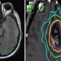
3.6 Brain
Reirradiation of brain tumours is often performed on an individual case-by-case basis and not yet based on the highest level of evidence (Veninga et al. 2001; Schwartz et al. 2015). In fact, no large randomised phase 3 trials have been published so far. It seems, however, that the selection criteria employed by experienced clinicians allow for retreatment, e.g. of glioblastoma with fractionated stereotactic radiotherapy, with low to moderate risk of late toxicity and encouraging median survival (Nieder et al. 2006b, 2008; Fogh et al. 2010). True long-term survival after reirradiation of brain tumours is unlikely and differentiation between tumour- and treatment-related deficits might be challenging. Therefore the actual rates of toxicity are difficult to estimate (Mayer and Sminia 2008; Sminia and Mayer 2012). In fact, toxicity risk might differ between brain regions with highly collateralised blood supply and those perfused by only few small branches. Importantly and in contrast to spinal cord and other organs, no animal experiments on reirradiation of the brain are available. Flickinger et al. (1989) published a series of nine patients with suprasellar or pituitary tumours having more than 5 years of follow-up after receiving two series of radiotherapy. Doses varied between 36 and 54 Gy for the first and 35–50 Gy for the second course. The median interval was as long as 7.5 years. The cumulative dose varied from 70 to 89 Gy in 2-Gy fractions with three patients receiving at least 86 Gy. Only one patient who was treated to 86 Gy developed a severe complication, namely, optic neuropathy. Of 32 patients treated by Dritschilo et al. (1981), 11 were alive at the time of analysis. Eight were experiencing productive lives and three were suffering from severe neurological damage including two cases of radionecrosis. Temporal lobe injury after treatment of nasopharynx cancer is not uncommon (Liu et al. 2014), and more details about this topic can be found in the chapter reviewing this particular tumour type.
Reirradiation of glioma with fractionated stereotactic radiotherapy (5 fractions per week, 5 Gy per fraction) after a median dose of 55 Gy was reported by Shepherd et al. (1997). All patients treated with more than 40 Gy developed late toxicity whereas only 25 % of those who received 30–40 Gy developed such adverse events. Several institutions have used reirradiation doses of 30–40 Gy with fraction sizes below 4 Gy with acceptable rates of toxicity (Combs et al. 2005; Fogh et al. 2010), and there is now also a prospective trial in progress (Combs et al. 2010). Radiosurgery was systematically studied by the RTOG (Shaw et al. 1996, 2000). Adults with cerebral or cerebellar solitary non-brainstem tumours ≤40 mm in maximum diameter were eligible. Initial radiosurgical doses were 18 Gy for tumours ≤20 mm, 15 Gy for that 21–30 mm and 12 Gy for that 31–40 mm in maximum diameter. Dose was prescribed to the 50–90 % isodose line. Doses were escalated in 3-Gy increments providing the incidence of irreversible grade 3 or any grade 4 or 5 RTOG central nervous system toxicity was <20 % within 3 months of radiosurgery. Between 1990 and 1994, 156 analysable patients were entered, 36 % of whom had recurrent primary brain tumours (median prior dose 60 Gy) and 64 % recurrent brain metastases (median prior dose 30 Gy). The median interval was 11 months, minimum 3 months. The maximum tolerated doses were 24 Gy, 18 Gy and 15 Gy for tumours ≤20 mm, 21–30 mm and 31–40 mm in maximum diameter, respectively. However, for tumours <20 mm, investigators’ reluctance to escalate to 27 Gy, rather than excessive toxicity, determined the maximum tolerated dose. The actuarial incidence of radionecrosis was 5 %, 8 %, 9 % and 11 % at 6, 12, 18 and 24 months following radiosurgery, respectively. Unacceptable toxicity was more likely in patients with larger tumours. Sneed et al. (2015) reported that radiosurgery followed by a second radiosurgery for recurrent brain metastases resulted in a 20 % 1-year cumulative incidence of symptomatic adverse radiation effect (median reirradiation dose 18 Gy, n = 72, crude risk 14 %). Compared to patients who had not received prior radiosurgery to the same lesion, the hazard ratio for any adverse radiation effect was 5.05 (95 % confidence interval 1.9–13.2).
3.7 Heart
Wondergem et al. (1996) assessed reirradiation tolerance of the heart at intervals of up to 9 months in a rat model by measuring cardiac function ex vivo 6 months after reirradiation. The cardiac tolerance dose was arbitrarily defined as the dose causing at least a 50 % function loss in one-half of the treated animals (ED50). Up to a 6-month interval, the reirradiation ED50 was close to the single-course ED50 but dropped significantly when the interval was longer than 6 months. The reirradiation tolerance also decreased with an increasing priming dose. A slow progression of damage induced by the priming dose is a plausible explanation for the decrease in tolerance with increasing time from the initial course. Systematic clinical data are not available.
3.8 Bladder
Stewart et al. (1990) studied whole bladder irradiation in mice. Priming doses were 8 Gy, which did not induce any observable functional bladder damage, and 16 Gy, which corresponded to 60 % of a full tolerance dose and produced a moderate level of damage starting from about 30 weeks. Reirradiation was given after approximately 13 or 39 weeks. Acute toxicity, manifesting as increased urination frequency, was mild when reirradiation was delivered 13 weeks after both priming dose levels but was amplified in the group receiving reirradiation 39 weeks after a priming dose of 16 Gy. However, with respect to late bladder damage (end point defined as >50 % reduction in bladder volume, increased urination frequency at ≥27 weeks after reirradiation), no long-term recovery was observed in up to a 9-month interval. Unfortunately, neither experimental data for partial organ reirradiation nor larger clinical studies on bladder re-exposure are available.
3.9 Kidney
Studies on mouse, rat and pig kidney suggested that radiation-induced toxicity progresses rather than recovers with time (Robbins et al. 1991; Stewart et al. 1994). For example, mouse experiments performed by Stewart et al. (1994) showed that reirradiation tolerance was inversely related to the priming dose, and tolerance decreased rather than increased significantly with increasing time interval. Analysis of the data on reirradiation at 26 weeks after lower priming doses using the linear-quadratic model gave an alpha/beta value of 1.4 Gy, which was significantly lower than the 3.3 Gy obtained for a single radiation course, indicating a larger fractionation-sparing effect for the reirradiation setting. Of clinical interest is also the finding in a rat model that single-dose cisplatin (5 mg/kg) administered 1 year after unilateral kidney irradiation progressively deteriorated the function of the irradiated kidney measured at 5 and 11 weeks after drug treatment (Landuyt et al. 1988). Compared with the contralateral kidney, the irradiated kidney showed a larger sensitivity to cisplatin-induced injury, even at subclinical doses of both drug and radiation.
4 Liver
The increasing numbers of patients with primary and secondary liver tumours receiving radiotherapy have led to occasional situations where the feasibility and potential of reirradiation for in-field or marginal progression or adjacent new lesions has to be discussed. Data are still very limited. Lee et al. (2016) treated 12 patients with hepatocellular carcinoma (median initial dose 50 Gy, range 36–60; median dose per fraction 1.8 Gy). Minimum interval to reirradiation was 5 months (median 20). The median dose was 50 Gy, range 36–58, with a median dose per fraction of 2.5 Gy. They aimed at a mean remaining liver dose ≤30 Gy with at least 700 cc of remaining liver volume. After reirradiation, 6 of 12 patients showed deterioration of liver function, and severe worsening of liver function (Child-Pugh score elevation ≥3 points) was also observed in four patients. The causal relationship was difficult to judge because several other local and systemic therapies were administered in most cases.
5 Discussion
Reirradiation should only be considered if the radiation tolerance of critical tissues or organs has not been exceeded during the first treatment, i.e. after careful review of the initial dose distribution and fields and assessment of organ status and side effects after the first treatment. Moreover, reasonable performance status and absence of other, less toxic treatment alternatives are prerequisites. It is important to weigh the expected benefits against morbidity and compromised quality of life. Solid preclinical evidence of recovery from occult radiation injury has been obtained for a variety of end points in several tissues and organs. However, these animal experiments cannot completely resemble the clinical situation, e.g. with regard to complexity of host factors. Cancer patients vary in biological and chronological age, cardiovascular and other comorbidity, organ reserve capacity and extent of multimodal therapies (additional surgery or even repeat surgeries, different types of anticancer drugs). It is also challenging to achieve dose distributions in partial organ irradiation of laboratory animals that are comparable to human IMRT and stereotactic RT. The majority of animal experiments were performed before such technology became available. It is therefore of utmost importance to collect clinical data on reirradiation tolerance. For many tissues and organs, an increasing body of data is now becoming available. Nevertheless, little is known for a number of end points such as neurocognitive function, damage to endocrine organs or the reirradiation tolerance of paediatric patients. Acutely responding tissues, in general, recover radiation changes practically completely within a few months and, therefore, can tolerate a repeat treatment course. For late injury end points, the heart, bladder and kidney did not exhibit long-term recovery (Fig. 2), whereas the skin, mucosa and spinal cord did. However, long-term recovery occurs within a defined time period that depends on the size of the priming dose and differs among tissues, species and age. Importantly, clinical evidence suggests that fibrosis, impaired blood perfusion and, in general, late normal tissue injury in humans continues to progress for many years and even decades. Lee et al. (1992) analysed more than 4,500 patients irradiated for nasopharyngeal cancer and found that the incidence of all types of late radiation toxicity, except of myelopathy, continued to rise even more than 10 years after treatment. Of course, individual prediction of toxicity risk would be a major step towards optimal choice of treatment intensity. Several reviews have summarised the current research efforts in this field (Coates et al. 2015; Jentsch et al. 2015; Kerns et al. 2014, 2015), which will not be discussed in any detail in this overview on reirradiation. The following book chapters provide both literature synopsis and exemplary cases that hopefully guide the choice of reirradiation regimens with acceptable complication probabilities. The paucity of data for many scenarios clinicians might face should encourage our community to embark on additional prospective clinical trials.
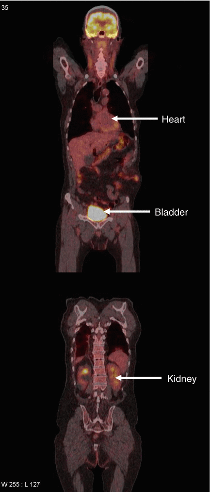
Fig. 2
Animal experiments suggest that radiation-induced toxicity progresses rather than recovers with time in the organs indicated by arrows (heart, kidney, bladder). Therefore, reirradiation of these organs might cause considerable late side effects
References
Abusaris H, Storchi PR, Brandwijk RP et al (2011) Second re-irradiation: efficacy, dose and toxicity in patients who received three courses of radiotherapy with overlapping fields. Radiother Oncol 99:235–239CrossRefPubMed
Related posts:

Stay updated, free articles. Join our Telegram channel

Full access? Get Clinical Tree


