(1)
Department of Radiology, John H Stroger Jr Hospital of Cook County, Chicago, IL, USA
Chest Wall Injury
Diagnosis
Flail Chest
Imaging Findings
1.
Fracture of the ribs at two or more sites of two or more contiguous ribs
2.
Parallel rows of the ribs in chest radiograph
3.
Adjacent pulmonary contusion, pneumothorax, and chest wall emphysema
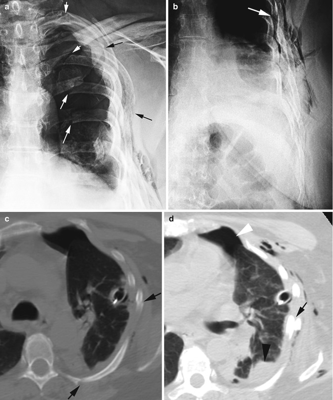
Fig. 7.1
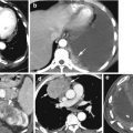
Flail chest with history of local trauma. (a, b) PA Chest x-rays show segmental fractures (white arrows show posterior fractures, black arrows show lateral fractures in a) of multiple ribs with displaced fractured segments forming two parallel rows of the ribs (arrow in b). (c) Axial CT shows two fractures of the same rib (arrows) and deformity of the thoracic wall due to the displacement of the segmental fractures. (d) Axial CT shows pneumothorax (white arrowhead), pulmonary contusion (black arrowhead), rib fracture (arrow), and emphysema of the thoracic wall
Mediastinal Injury
Diagnosis
Pneumomediastinum
Imaging Features
1.
Linear air parallel to the heart border
2.
Continuous diaphragm sign
3.
Air around the pulmonary artery and main branches—ring around artery sign
4.
Air outlining the major aortic branches—tubular artery sign
5.
Air outlining the bronchial wall—double bronchial wall sign
6.
Elevated thymus—thymic wing sign
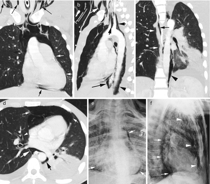
Fig. 7.2


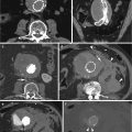
Pneumomediastinum. History of trauma. (a) Coronal reformatted CT shows air parallel to the left heart border (white arrows) and diaphragmatic surface of the heart (short black arrow). Gas is also seen tracking from the superior mediastinum to the neck around the vessels (long black arrows). (b) Sagittal reformatted image shows air around the distal esophagus (long black arrow) and adjacent aorta (black arrowhead). Air tracking to the neck around the aortic branches (short black arrows) and fascial planes (white arrow). (c) Coronal reformatted image shows air around the azygos vein (arrows) and around the descending aorta (arrowhead). (d) Axial CT shows air in the anterior mediastinum causing mild compression of the surrounding lung (thick white arrows). Air is seen surrounding the right lower lobe pulmonary artery (thin white arrow) and bronchus (arrowhead). Air is also surrounding the esophagus and aorta (black arrows). (e) PA chest x-ray shows linear air surrounding the heart and extending above the arch of the aorta (arrows). (f) Lateral chest x-ray shows air in the anterior mediastinum (arrowheads) and around the esophagus (arrows)
Stay updated, free articles. Join our Telegram channel

Full access? Get Clinical Tree


