Ulcerative Colitis

 Terminal ileum affected in minority of patients


• Toxic megacolon
image Colon is dilated with loss of fold and mucosal pattern

image Ascites is common ± pneumatosis, pneumoperitoneum


TOP DIFFERENTIAL DIAGNOSES


• Granulomatous colitis (Crohn disease)

• Infectious (including Clostridium difficile) colitis
image D egree of mucosal enhancement  and  submucosal edema is usually greater than seen in ulcerative colitis

• Ischemic colitis
image Rectum is almost always spared in ischemic colitis

• Cathartic colon

• Neutropenic enterocolitis

• Diverticulitis


PATHOLOGY


• Associated pathology
image Greater risk of colorectal cancer in UC than Crohn colitis
– Multiple carcinomas in 25% of UC cases

image Primary sclerosing cholangitis, uveitis

image Ankylosing spondylitis, rheumatoid arthritis


CLINICAL ISSUES


• Most common signs/symptoms
image Relapsing bloody mucus diarrhea

image Fever, weight loss, abdominal pain and cramps

• Initial onset: 15-25 years (small peak at 55-65 years)

• Begins in rectum with proximal continuous extension to part or all of colon


DIAGNOSTIC CHECKLIST


• Consider UC in any patient with sclerosing cholangitis

• Consider other causes of colitis, especially infectious and Crohn disease

image
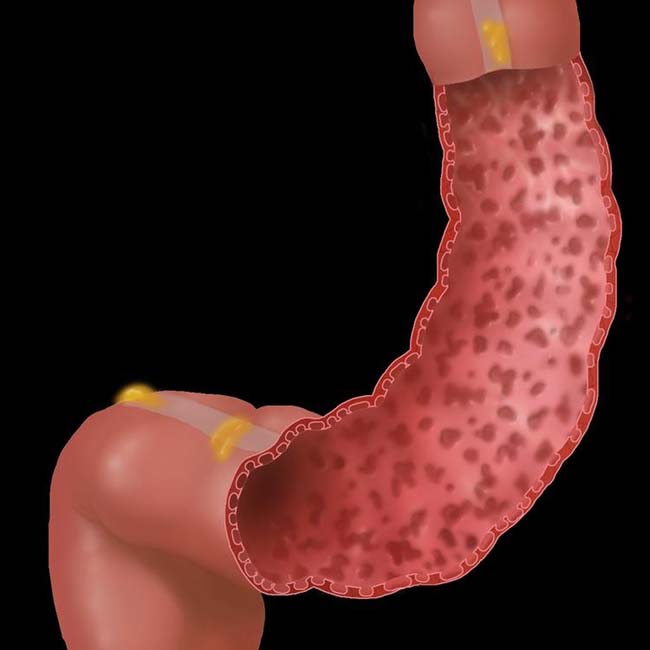
(Left) Graphic illustration demonstrates innumerable “collar button” ulcers and a loss of haustra throughout the descending and sigmoid colon.


image
(Right) Single-contrast barium enema shows innumerable “collar button” ulcers image 
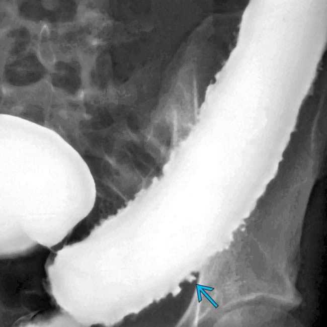
and loss of haustra throughout the descending colon.

image
(Left) This 51-year-old woman has an acute flare of chronic ulcerative colitis. Coronal CECT shows pancolitis with mucosal hyperenhancement and submucosal edema image
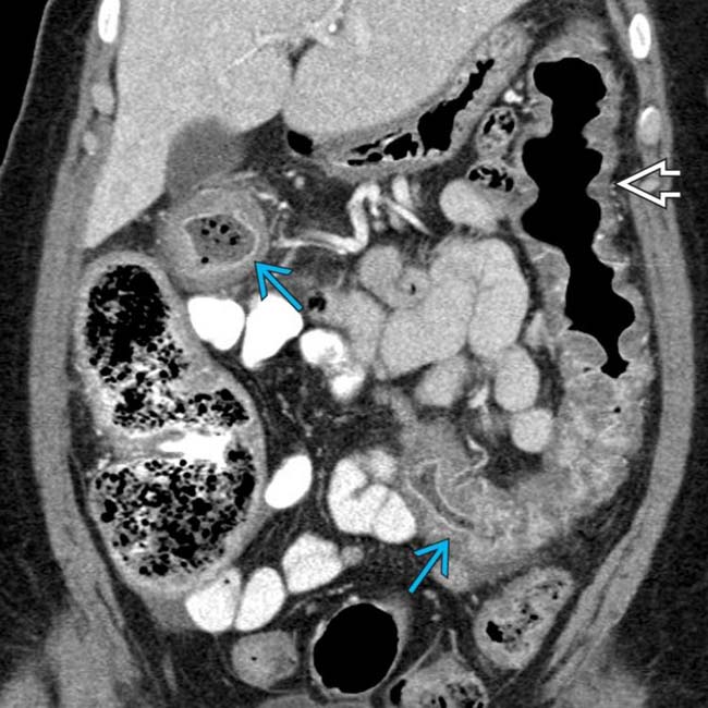
, with blunted transverse folds image.


image
(Right) Axial CT in the same patient shows the mucosal hyperenhancement and submucosal edema image
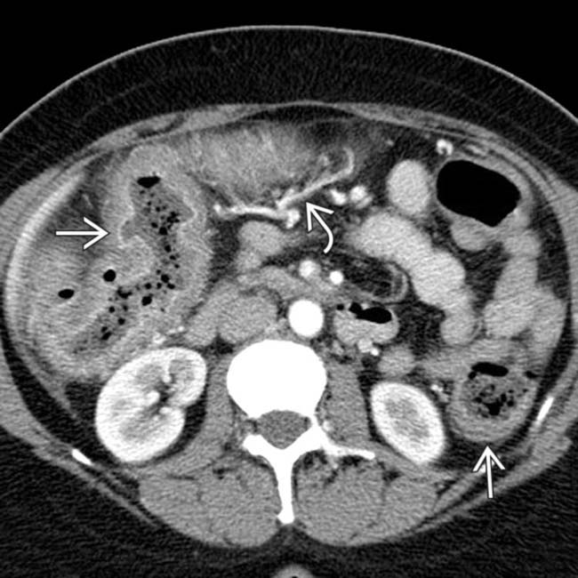
. Note the prominent vessels image supplying the inflamed colon.


TERMINOLOGY


Abbreviations


• Ulcerative colitis (UC)


Definitions


• Chronic, idiopathic, diffuse, inflammatory disease primarily involving colorectal mucosa


IMAGING


General Features


• Best diagnostic clue
image Distal or pancolitis with mucosal hyperenhancement and only moderate submucosal edema

• Location
image Rectum only (30%), rectum and distal colon colon (40%), pancolitis (30%)
– Terminal ileum affected in minority of patients

• Morphology
image Moderate wall thickening and luminal narrowing in acute phase

image Foreshortened and ahaustral colon in chronic phase
– Described as “lead pipe” or “windowpane”


Fluoroscopic Findings


• Barium enema
image Acute
– Colorectal narrowing, incomplete filling (spasm + irritability)

– Fine mucosal granular pattern (edema/hyperemia)

– Mucosal stippling: Punctate barium collections and ulcers due to mucosal erosion and crypt abscesses

– Flask-shaped “collar button” ulcers 
image Ulcers may progress to widespread mucosal sloughing

image Residual or hyperplastic mucosa may appear as pseudopolyps

– Thickened transverse folds due to submucosal edema

image Chronic
– Shortened, less redundant colon

– “Lead pipe” or “windowpane” colon

– Blunted or complete loss of transverse folds

– Luminal narrowing and widened presacral space > 1.5 cm

– Benign or malignant strictures


CT Findings


• CECT
image Target or halo sign
– Enhancing inner ring of bowel wall (mucosa)

– Nonenhancing middle ring of bowel wall (submucosa)

– Enhancing outer ring of bowel wall (muscularis propria and serosa)

image Enhancement of mucosal islands or inflammatory pseudopolyps

image Wall thickening generally < 10 mm

image Perirectal fibrofatty proliferation and narrowing of rectal lumen
– Results in widening of presacral (retrorectal) space

image Toxic megacolon
– Colon is dilated, often > 8 cm (on CT, more on plain films)
image Small bowel also dilated (ileus)

– Colonic wall may be thick or thin

– Loss of normal transverse folds and mucosal pattern
image Mucosal islands or pseudopolyps may be seen

– Ascites is common ± pneumatosis, pneumoperitoneum


Imaging Recommendations


• Best imaging tool
image CECT with multiplanar reformations

image Air-contrast barium enema provides excellent depiction of colonic mucosal disease
– Rarely requested or performed in era of easy access to CT and colonoscopy


DIFFERENTIAL DIAGNOSIS


Granulomatous Colitis (Crohn Disease)


• Granulomatous colitis (GC) is transmural inflammation while UC is generally more limited to mucosa

• More often has skip lesions and involvement of small bowel

• Barium enema
image Aphthous ulcers: Shallow collection of barium in mucosal ulceration with surrounding edema

image “Cobblestoning”: Longitudinal and transverse ulcerations with paving stone appearance

image Segmental distribution: Colon and small bowel (60% of cases); isolated to colon (20% of cases)

Only gold members can continue reading. Log In or Register to continue

Stay updated, free articles. Join our Telegram channel

Nov 16, 2016 | Posted by in GASTROINTESTINAL IMAGING | Comments Off on Ulcerative Colitis

Full access? Get Clinical Tree

Get Clinical Tree app for offline access