13 Ultrasound-Guided Shoulder Injections
♦ Long Head of the Biceps
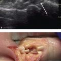
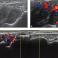
• The biceps is first located in the longitudinal axis between the lesser and greater tuberosities (Fig. 13.1a,b).
• The most proximal aspect of the tendon is then identified between the tuberosities.
• The area just superior to the probe should be prepped.
• The needle should be inserted slowly in line with the probe angled toward the biceps tendon (Fig. 13.1c).
• When the tip of the needle is visualized within the bicipital sheath, injection can proceed.

Fig. 13.1 (a,b) The long head of the biceps viewed in the short axis. (c) Injection into the long head of the biceps.
♦ Subacromial
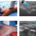
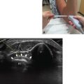
• The patient should place the hand on ipsilateral hip and adduct the shoulder(Fig. 13.2a,b).
• The anterior aspect of the supraspinatus should be visualized in the longitudinal plane.
• Scanning proceeds slightly anteriorly to locate the biceps tendon and ensure the proper position has been attained.
• The probe is then returned to the previous position, and the area just lateral to the probe is prepped.
• The needle should be inserted slowly from a lateral to medial direction in line with the probe until it is visualized just superficial to the supraspinatus (Fig. 13.2c).