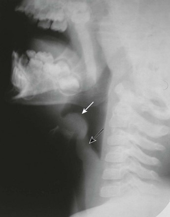
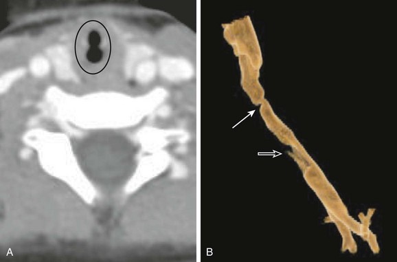
Chapter 51 Upper airway disease is a common problem in the pediatric age group. Affected children usually present with acute respiratory distress associated with stridor, apnea, or even acute pulmonary edema. Some children may present with a more chronic course of clinical symptoms characterized by recurrent chest infections or obstructive sleep apnea, which can lead to growth retardation, chronic respiratory failure, cor pulmonale, and even death.1 Children with upper airway obstruction usually present with stridor, a harsh respiratory noise caused by turbulent air passing through a narrowed airway. Timing of stridor in relation to the respiratory cycle can indicate the location of the narrowing. Inspiratory stridor usually results from obstruction above the level of the glottis, whereas expiratory stridor is characteristic of intrathoracic obstructions. Biphasic stridor suggests obstruction in the area between the glottis and subglottis or fixed/critical obstruction at any level.2 Although a thorough history and physical examination may narrow differential diagnostic considerations of upper airway disease in the pediatric population, imaging evaluation plays an important role in establishing the diagnosis, localizing the anatomic area of abnormality, and defining the extent of disease. Frontal and lateral radiographs of the airway and fluoroscopy have been mainstays in upper airway evaluation. Advances in cross-sectional imaging techniques approaching near endoscopic detail have been developed to more accurately demonstrate airway anatomy.3 For practical purposes, the discussion of pathology in this chapter is divided into the supraglottic, glottic, and subglottic regions. Etiology: Laryngomalacia is abnormal laxity of the pharyngeal tissues that causes the epiglottis, arytenoids, and aryepiglottic folds to involute and partially obstruct breathing during inspiration. The underlying cause of laryngomalacia is attributed to the immaturity of the laryngeal cartilages and muscles, which allows the larynx and the supralaryngeal structures to collapse.1–4 Laryngomalacia accounts for more than 75% of cases of congenital stridor and is the most common cause of symptomatic partial upper airway obstruction in infants.5 The stridor typically improves when the infant is agitated or active and becomes worse when the infant is at rest. Imaging: The diagnosis of laryngomalacia can be established with use of airway fluoroscopy, although laryngoscopy is the diagnostic procedure of choice.6 The characteristic imaging finding in patients with laryngomalacia consists of downward and posterior bending of the epiglottis and anterior buckling of the aryepiglottic folds, which narrow and eventually obliterate the upper airway (Fig. 51-1). Although airway fluoroscopy appears to be reliable because of its high specificity, it has low sensitivity, and thus negative findings of a fluoroscopic study require further diagnostic evaluation in the setting of a high clinical suspicion of laryngomalacia.7 Figure 51-1 Laryngomalacia in a 3-month-old boy with stridor. Treatment and Follow-up: In most infants with laryngomalacia, stridor resolves during the first year of life. However, potentially serious complications, including airway obstruction and sudden death, also may occur. In severe or life-threatening cases, the following surgical treatment options currently are available: supraglottoplasty, an incision in the aryepiglottic folds, epiglottopexy, and tracheostomy.8 Etiology: Acute bacterial epiglottitis is a potentially serious cause of upper airway obstruction in children. It is characterized by inflammation and swelling of the epiglottis and the aryepiglottic folds but also can affect the false cords and the subglottic region.9 Epiglottitis usually is attributed to Haemophilus influenzae, although other bacteria including Streptococcus, Staphylococcus, Moraxella, and Pseudomonas also have been implicated.10 Because Haemophilus influenzae is now preventable by immunization, the incidence of epiglottitis has substantially diminished, although vaccine failures do occur.11 Epiglottitis typically occurs in preschool children between 3 and 6 years of age, but it also can occur in adults.12 Epiglottitis usually has a sudden onset with no history of a preceding upper respiratory tract infection. Affected children typically appear toxic with acute stridor, dysphagia, fever, restlessness, drooling, and increased respiratory distress in the recumbent position. Epiglottitis is a life-threatening disease that requires potential emergent intubation. Imaging: Epiglottitis usually is diagnosed with radiography, and endoscopy is obtained for confirmation. A lateral radiograph reveals swelling and marked enlargement of the epiglottis that resembles the shape of a thumb (Fig. 51-2). Associated thickening of the aryepiglottic folds also occurs. Inflammation extends into the glottis and subglottic regions, causing a steeple or funnel configuration of the airway on frontal views. Because of the potentially rapid progression of the condition, which can lead to sudden and complete obstruction of the upper airway, radiographs should be obtained expeditiously with minimal manipulation of the neck. When undergoing imaging, patients should remain upright in their position of maximal comfort. Conditions that mimic bacterial epiglottitis in children include caustic ingestion, angioneurotic edema, chemical or thermal injury, an abscess, and epithelial cyst, among others. The omega epiglottis with prominent lateral folds is a normal variant and should not be misinterpreted as epiglottitis. In such cases, the aryepiglottic folds remain thin.13 Etiology: Laryngeal atresia, a rare congenital malformation, is usually fatal. The malformation is caused by nondevelopment of the sixth branchial arch during embryogenesis, resulting in failure of the larynx and the trachea to recanalize. Typical presentation of affected patients with laryngeal atresia at birth is severe respiratory distress despite strong respiratory effort. Associated anomalies include a tracheoesophageal fistula, esophageal atresia, urinary tract abnormalities, limb defects, and low-set ears.14 Imaging: Laryngeal atresia can be diagnosed with use of prenatal ultrasound by identifying the signs of congenital high airway obstruction syndrome, such as hyperechogenic lungs, a flattened or inverted diaphragm, a dilated and fluid-filled trachea, fetal hydrops, and polyhydramnios.15 Fetal magnetic resonance imaging (MRI), which correlates highly with prenatal ultrasound, can detect laryngeal atresia with the advantage of having a capability to identify the level of obstruction in most cases (Fig. 51-3).16 Figure 51-3 Congenital high airway obstruction syndrome in a 20-week-gestation fetus. Treatment and Follow-up: The treatment of laryngeal atresia focuses on prompt airway intervention at delivery after an accurate prenatal diagnosis, which may allow survival of infants with this otherwise fatal condition. An emergent tracheostomy is required immediately upon birth to secure an airway. Repair of laryngeal atresia requires laryngotracheal reconstruction. Etiology: A congenital laryngeal web is an uncommon condition caused by faulty embryogenesis of the laryngotracheal groove. This web is a band of varying thickness, usually located anteriorly at the level of the cords, which obliterates the anterior commissure. A congenital laryngeal web occasionally occurs just below the true cords. Affected infants present with a weak or absent cry, varying degrees of stridor, and respiratory distress, and they often may have concomitant abnormalities such as subglottic stenosis.1 Imaging: A definitive diagnosis of laryngeal web can be provided by direct laryngoscopy, but imaging evaluation plays a role in diagnosis. Plain radiography and fluoroscopy can demonstrate that the abnormality is in the glottis region, but no definitive radiographic findings exist.5 Computed tomography (CT) with multiplanar imaging and three-dimensional reconstructions can provide precise information with respect to the exact location of the web, its thickness, and its extent (Fig. 51-4). Furthermore, CT is able to visualize the structures beyond the area of obstruction, which is a limitation of laryngoscopy.17 Figure 51-4 Laryngeal web. Treatment and Follow-up: The primary goals of treatment for a congenital laryngeal web are to provide a patent airway and achieve a sufficient voice quality. Treatment depends on the thickness and extent of the abnormality. Surgical procedures include laryngotracheal reconstruction, laryngofissure and placement of a stent or keel, and endoscopic lysis with application of mitomycin C.18,19 Etiology: A laryngocele is an abnormal dilatation of the laryngeal saccule, which is a narrow blind pouch arising from the anterior end of the laryngeal ventricle, extending superiorly into the paralaryngeal space, and bounded laterally by the thyroid cartilage.20 A laryngocele may be congenital or acquired. Laryngoceles occur more often in males than in females. In neonates and young children, a laryngocele is likely congenital and is contingent upon the presence of a saccular dilatation of the ventricular appendage. Increased laryngeal pressure may cause the sac to distend, at times extending into the aryepiglottic fold.21 A laryngocele is classified as internal if it lies within the larynx and external if it protrudes through the thyrohyoid membrane. The most common type is mixed. As the laryngocele communicates with the larynx, it normally contains air but may be filled with mucus or fluid.22 An acquired laryngocele may develop in older children or adults who experience increased intralaryngeal pressure, such as glass blowers, persons who play wind instruments, or persons with a chronic cough. Imaging: In the past, anteroposterior (AP) and lateral soft tissue neck radiographs, linear tomography, and contrast laryngoscopy were used to identify laryngoceles.21 However, CT and/or MRI have replaced those older imaging techniques in recent years.20 On CT or MRI, a laryngocele typically is a well-defined structure of air or near-water density value in a characteristic location, with a smooth surface and lack of mucosal abnormality (Fig. 51-5). The exact location and extent of upper airway narrowing due to a laryngocele also can be well evaluated with CT or MRI. Figure 51-5 A laryngocele in a 5-year-old boy with chronic stridor. Etiology: Recurrent respiratory papillomatosis (RRP) is the most common benign neoplasm to affect the larynx in children.23 It is the proliferation of benign squamous papillomas in the aerodigestive tract, usually involving the larynx. RRP tends to recur, spreads throughout the aerodigestive tract, and can even undergo malignant transformation. The etiology of RRP is infection of the upper airway with human papillomavirus types 6 and 11.24 Vertical transmission occurring during delivery through an infected birth canal is presumed to be the major mode of transmission in children.25 Change in voice (e.g., hoarseness, weak cry, and aphonia) and stridor are the most common symptoms in patients with RRP. Less common presenting symptoms include chronic cough, recurrent pneumonia, failure to thrive, dyspnea, dysphagia, and acute respiratory distress, especially in infants with an upper respiratory tract infection.26 The age of symptom onset typically ranges from neonates to 6 years. RRP can mimic other diseases including croup, tracheomalacia, or asthma. This diagnosis should be considered in children when other common pediatric airway diseases either do not follow the expected natural history or do not respond to treatment.27 Imaging: On plain radiographs, RRP typically appears as an irregular filling defect in the glottis (Fig. 51-6). It also can extend into the subglottic region and seed peripherally in the respiratory tract, resulting in pulmonary nodules or nonspecific lung parenchymal changes.1 A confirmatory diagnosis of RRP is made by direct laryngoscopy and biopsy for tissue diagnosis.27 Figure 51-6 Recurrent respiratory papillomatosis in a 4-year-old boy. Etiology: Subglottic stenosis is a narrowing of the subglottic airway that can be either congenital or acquired. In pediatric patients, congenital subglottic stenosis is due to faulty recanalization of the fetal laryngeal lumen, whereas the acquired type usually is the result of prolonged endotracheal intubation.1 Because of improved management of neonates who require ventilatory support, the incidence of neonatal subglottic stenosis has diminished substantially.28 Stridor is the most common presentation. However, an infant or child with a mild degree of congenital or acquired subglottic stenosis may remain asymptomatic until an upper respiratory tract infection results in aggravation of the subglottic airway narrowing.4 Imaging: Endoscopy is the reference standard for diagnosis, but imaging plays an important role in the evaluation of subglottic stenosis. Plain radiograph and fluoroscopy, which show fixed narrowing of the subglottic airway, usually enable diagnosis. However, CT with virtual bronchoscopy and MRI can be useful in assessing the exact site and length of stenosis and the airway distal to the area of narrowing (Fig. 51-7).4,29
Upper Airway Disease
Supraglottic Abnormalities
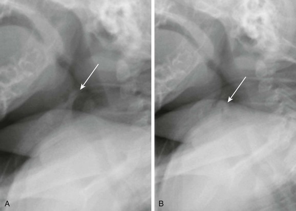
A, Still image from an airway fluoroscopic study demonstrates the normal position of the epiglottis (arrow). B, Still image from an airway fluoroscopic study shows laxity of the epiglottis with posterior, downward movement obstructing the airway (arrow) consistent with laryngomalacia.
Epiglottitis
Glottic Abnormalities
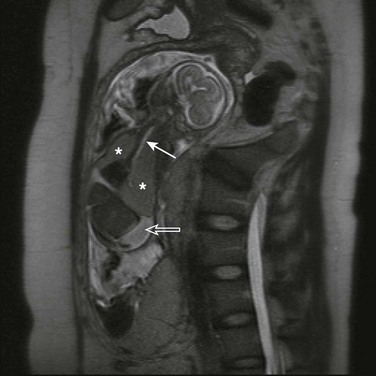
A half Fourier acquisition single-shot turbo spin echo sequence of a fetal magnetic resonance imaging scan demonstrates a dilated, fluid-filled trachea up to the mid neck (solid arrow), enlarged hyperintense lungs (asterisks) with inversion of both hemidiaphragms, and ascites (open arrow). (Courtesy of Andrew Mong, MD, Philadelphia, PA.)
Laryngeal Web
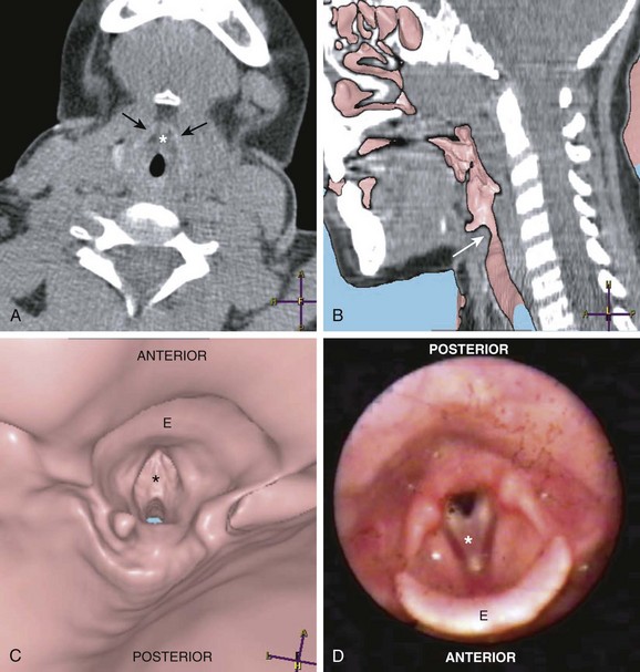
A, An axial computed tomography (CT) scan of the neck at the level of the larynx and the thyroid cartilage (black arrows) demonstrates the laryngeal web (asterisk). B, A sagittal virtual endoscopic view superimposed on a sagittal reformatted CT image shows the laryngeal web (arrow). A virtual endocsopic view (C) and corresponding larygoscopy (D) demonstrate the laryngeal web (asterisk). The epiglottis (E) is well demonstrated on these images. (Courtesy of Suleymen Men, MD, Izmir, Turkey.)
Laryngocele
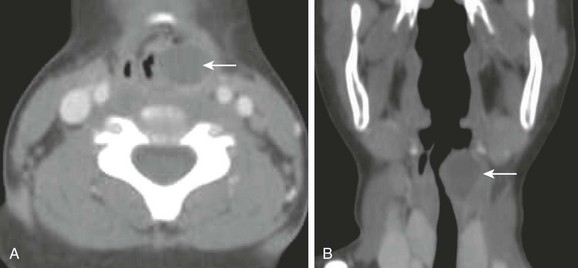
An axial computed tomography scan (A) with coronal reconstruction (B) of the upper airway reveals a well-demarcated fluid-filled rounded structure (arrow) adjacent to the left laryngeal ventricle compatible with a laryngocele. Also noted is a marked narrowing of the upper airway.
Recurrent Respiratory Papillomatosis
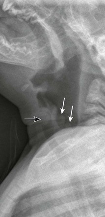
A lateral soft tissue radiograph demonstrates a rounded soft tissue mass in the laryngeal region (black arrow). Nodular soft tissue densities also are noted along the aryepiglottic folds (white arrows). (Courtesy Elizabeth H. Ey, MD, Dayton, OH.)
Subglottic Abnormalities