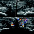
Fig. 19.1
Posttraumatic pseudoaneurysm of a digital artery. The color Doppler examination reveals permanent dilatation of the artery with rotatory flow within the aneurysm (arrow) (a–c)
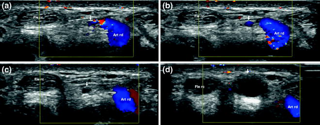
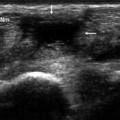
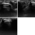
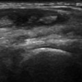
Fig. 19.2
Posttraumatic pseudoaneurysm of a radial artery branch in the thenar eminence. The sonogram reveals a small arterial branch (arrows) that runs from the ulnar side of the wrist toward the flexor carpi radialis (Fle rc) and connects the lesion to the radial artery (Art rd) (a–d). The scan also documents the location, size, and thrombosed nature of the aneurysm (arrow) © (in d)
Trauma to the latter region, for example, can easily damage the superficial branch of the ulnar artery at its emergence from the Guyon’s canal. At this level, the course of the artery is fairly superficial, and it protected only by the palmaris brevis and subcutaneous muscles. As a result, traumatic compression of this region often crushes the artery against the hook of the hamate. In the thenar eminence, the deep palmar arch is similarly vulnerable to trauma at the point where it crosses the second metacarpal.
Aneurysms can develop in any of the arteries of the hand. However, they are especially common in the superficial branch of the ulnar artery and the palmar branch of the radial artery. Repeated blunt trauma to the palm of the hand compresses the former vessel against the hook of the hamate, the latter against the tubercle of the trapezium [1–5].
The damage to the arterial wall produced by this trauma generally involves all three layers (intima, media, and adventitia) and may result in the formation of an aneurysm.
The clinical manifestations include soft tissue swelling and symptoms related to vasospasm, ischemia, and claudication [6].
On ultrasound, the artery appears dilated (Fig. 19.1) and often contains hypoechoic thrombotic material (Fig. 19.2), which can cause stenosis or complete occlusion of the lumen. Color Doppler imaging reveals reduction in the linear flow velocity and the presence of rotator flow [6] (Fig. 19.2).
The main complications of aneurysms involving the hand are thrombosis and distal embolism. Treatment is surgical and involves resection of the aneurysm with end-to-end anastomosis [3].
Atherosclerosis at the level of the hand has certain peculiar features: it is typically diagnosed in smokers or patients with chronic renal failure, and it is characterized by the absence of fibrocalcific plaques. It presents with pain followed later by ischemia involving the digits [7, 8].
The arteries are tortuous, rigid, and serpiginous—features that are clearly depicted on sonography. Color Doppler imaging demonstrates loss of laminar flow and the presence of turbulent flow (Fig. 19.3), and it can also be useful for identifying vessel obliteration.
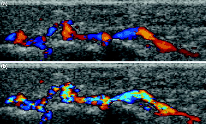
Fig. 19.3
Atherosclerosis of the hand. Sonography reveals tortuous, rigid, serpiginous arteries; color Doppler imaging demonstrates loss of laminar flow and the presence of turbulent flow (a, b)
As for hereditary hemorrhagic telangiectasia—also known as Osler-Weber-Rendu syndrome—it is an autosomal dominant disorder characterized by vascular dysplasia and a prevalence in the general population of 1–2 per 100,000. The syndrome is associated with telangiectasias and arteriovenous malformations involving skin, mucosa, and visceral tissues. The most common complications of mucosal involvement are epistaxis and gastrointestinal bleeding [9, 10]. Visceral involvement is also observed in the lungs and liver. In 90 % of all cases, the disease presents with epistaxis that occurs before the age of 21. It can lead to severe blood loss.
Telangiectasias also develop on the mucosal surface of the tongue (where hemorrhage is particularly difficult to control) or lips, face, conjunctiva, ears, and fingers. Involvement of the fingers and wrists is present in 40 % of all cases [10]. The alterations observed in the hands and wrists are similar to those seen in other parts of the body. They can involve all vascular structures, regardless of their size, and include dilatation and/or complex changes in arteries and veins, as well as arteriovenous fistulas (Fig. 19.4). Ultrasonography is a sensitive and specific method for detecting and identifying the lesions of Osler-Weber-Rendu syndrome.
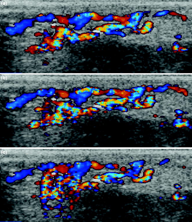
Fig. 19.4


Related posts:
Stay updated, free articles. Join our Telegram channel
Full access? Get Clinical Tree
