(1)
Department of Radiology, John H Stroger Jr Hospital of Cook County, Chicago, IL, USA
Kommerell Diverticulum
Diagnosis
Stenosis of Origin of Anomalous Left Subclavian Artery with Kommerell Diverticulum
Imaging Features
1.
Right-sided arch of the aorta
2.
Stenosis of aberrant left subclavian artery with post stenotic dilatation
3.
Kommerell diverticulum of the aorta at origin of the subclavian artery
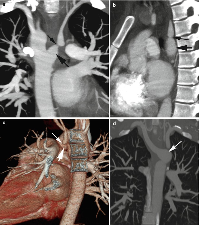
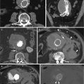
Fig. 6.1
Stenosis of the subclavian artery. Aberrant origin of left subclavian artery from right-sided arch of the aorta with Kommerell diverticulum at its origin and stenosis distal to the diverticulum presenting with subclavian steal syndrome. (a) Coronal reformatted image shows Kommerell diverticulum (thick arrow) at origin of the aberrant subclavian artery and marked stenosis distal to the diverticulum (thin arrow) with post stenotic dilatation. (b, c) Oblique sagittal reformatted image and 3D volume-rendered images show stenosis of left subclavian artery (thin arrows) and the Kommerell diverticulum (thick arrows). (d) Coronal reformatted image in different patients shows Kommerell diverticulum (arrow) at left-sided descending thoracic aorta at the origin of aberrant right subclavian artery
Coarctation of Aorta
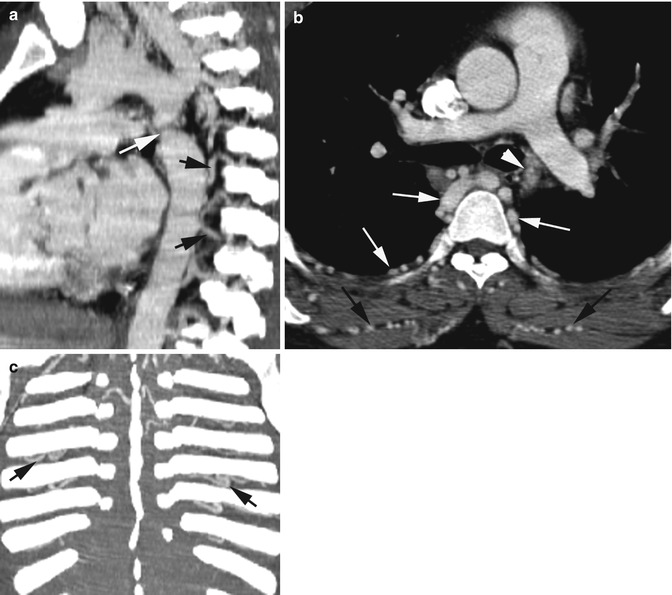
Fig. 6.2
Coarctation of the aorta in adult. (a) Sagittal reformatted image shows short segment of stenosis of the aorta in the region of the ductus (white arrow) with dilated intercostal arteries (black arrows). (b) Axial CT shows narrowed segment of the aorta (arrowhead), dilated intercostal arteries (white arrows), and also dilated intermuscular arteries. (c) Coronal reformatted image shows dilated intercostal arteries (arrows)
Renal Artery Stenosis
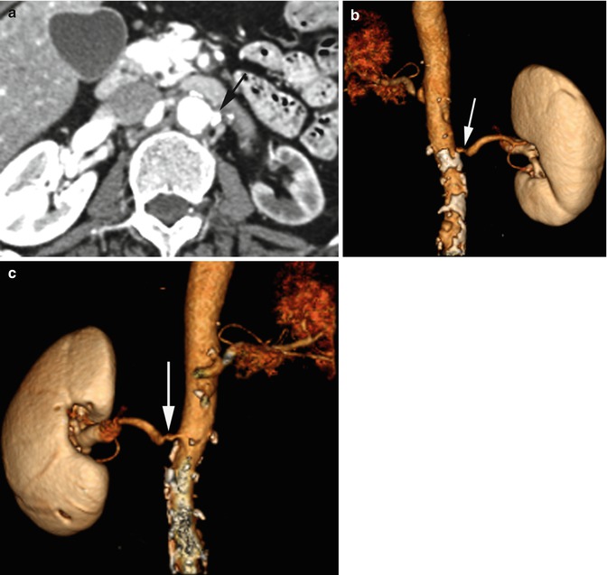
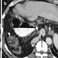
Fig. 6.3
Stenosis of the renal artery. Atherosclerosis with stenosis of the left renal artery. Contrast-enhanced axial CT (a) shows large calcification at origin of the left renal artery (arrow). The left kidney is atrophic with decreased perfusion. 3D volume-rendered images, (b) frontal view, and (c) posterior view show narrowing of origin of the left renal artery (arrow)
Mediastinal Fibrosis
Diagnosis
SVC Obstruction by Mediastinal Fibrosis
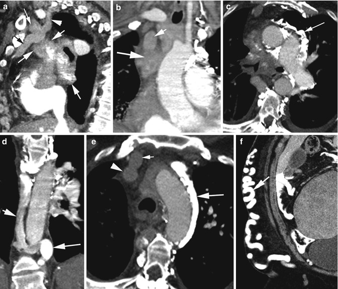
Fig. 6.4
Superior vena cava obstruction by mediastinal fibrosis confirmed by biopsy. Urine Blastomyces antigen was positive. CECT (a, b) sagittal, coronal reformatted, and (c) axial images show large mediastinal mass (thick arrows) with areas of calcification (long thin arrow in c) obstructing the SVC (short arrow in b) with dilatation of the internal thoracic vein (black arrow), IJ vein (thin arrow in a), and the intercostal vein (arrowhead). (d) Coronal reformatted CT shows dilated azygos (short arrow) and hemiazygos vein (long arrow). (e) Axial CT shows dilated left superior intercostal vein (long arrow), internal thoracic vein (short arrow), and SVC (arrowhead). (f) Dilated veins in the right abdominal wall subcutaneous tissue which drain into the femoral vein (arrow)
IVC Stenosis
Diagnosis
Tumor Compression of IVC and Hepatic Veins
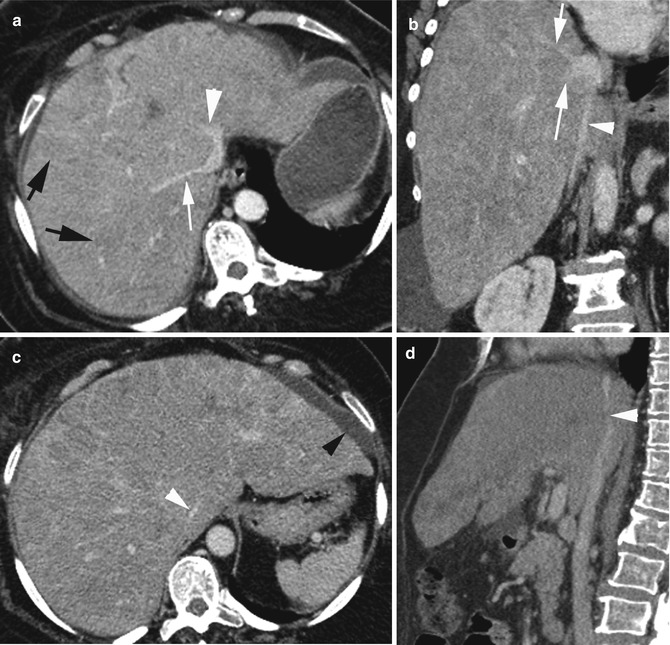
Fig. 6.5
Intrahepatic IVC and hepatic vein compression by tumor. Patient with infiltrating ductal breast cancer with diffuse liver metastasis. CECT (a) axial shows markedly compressed IVC, narrowed right (white arrow) and left hepatic veins (arrowhead) and absent middle hepatic vein (long arrow in b), and diffuse low-density metastasis (black arrows). (b, c, d) Coronal reformatted, axial, and sagittal reformatted CT show markedly narrowed IVC (white arrowhead) and small ascites (black arrowhead)
Diagnosis
Hypoplastic IVC
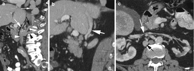
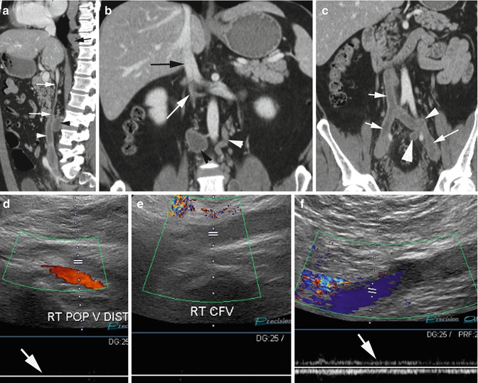
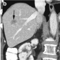
Fig. 6.6
Hypoplastic infrahepatic IVC, incidental finding. (a) Sagittal reformatted image shows narrow caliber of IVC (thick white arrow). The IVC below the renal veins is normal in size (arrowhead). Collateral flow is seen through the dilated ascending lumbar vein (black arrow) draining into the dilated azygos vein (thin white arrow) and the lumbar vein (black arrowhead). (b) Sagittal reformatted CT shows normal IVC (arrow) within the liver. (c) Axial CT shows very narrow IVC (white arrow) between the renal veins and dilated ascending lumbar vein (black arrow)
Fig. 6.7
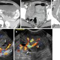
Hypoplastic IVC with thrombus. (a) Sagittal reformatted image shows a very narrow segment of IVC (arrows) below the renal veins. Thrombus distending the IVC below the stenotic region (white arrowhead) and thrombus in a lumbar vein (black arrowhead). (b) Coronal reformatted CT shows normal size of IVC at the renal veins and with thin median septa (black arrow). IVC narrow below the renal veins (white arrow). The left ascending lumbar vein (white arrowhead) is dilated and there is thrombus in a right lumbar vein (black arrowhead). (c) Coronal reformatted CT shows thrombus in IVC, iliac veins (white arrows), in the origin of ascending left lumbar vein (thin arrowhead), and median sacral vein (thick arrowhead). (d, e) Duplex and spectral Doppler imaging of the right leg shows thrombus in the right popliteal vein extending to CFV with absent spectral flow (arrow in d). (f) Left common femoral vein is patent but shows lack of respiratory variation due to proximal occlusion (arrow)
Bibliography
1.
2.
3.
4.
5.
6.
7.
8.
Castaner E, Alguersuari A, Gallardo X, et al. When to suspect pulmonary vasculitis: radiologic and clinical clues. Radiographics. 2010;30:33–53.CrossRefPubMed
Related posts:
Stay updated, free articles. Join our Telegram channel

Full access? Get Clinical Tree


