Hypervascular pancreatic mass with multiple peptic ulcers and thickened folds
• Best imaging tool
Helical CT or MR for pancreas and possible metastasis
Endoscopic ultrasonography for additional primary sites; guides biopsy
TOP DIFFERENTIAL DIAGNOSES
• Helicobacter pylori gastritis
• Gastric carcinoma
• Gastric metastases and lymphoma
• Extrinsic inflammation
• Other gastritides
PATHOLOGY
• 20-60% of cases are associated with multiple endocrine neoplasia type 1 (MEN1)
CLINICAL ISSUES
• Most common signs/symptoms
Pain, increased acidity, severe reflux, diarrhea, upper gastrointestinal tract ulcers
Gastrinomas are often multiple (60%), malignant (60%), and metastatic (30-50%)
• Hypergastrinemia is hallmark of Zollinger-Ellison syndrome (ZES)
Serum gastrin level of > 1,000 pg/mL is virtually diagnostic of ZES
• Prognosis
Good with surgical resection of primary gastrinoma
Poor if gastrinoma, liver metastases, or ulcers recur after surgery
(Left) Axial CECT in a 63-year-old man who presented with intractable peptic ulcer disease demonstrates hyperemia and mural thickening of the stomach.
(Right) Arterial phase CECT in the same patient shows a small hypervascular gastrinoma in the pancreatic head. It is important to distinguish this from the superior mesenteric artery and superior mesenteric vein .
(Left) Axial CT of a 55-year-old woman with hypercalcemia, diarrhea, and severe abdominal pain as presenting symptoms of MEN1 syndrome shows one of several neck masses , representing parathyroid adenomas or hyperplasia.
(Right) Abdominal CT in the same case shows marked hypervascularity and thickening of the gastric wall . Multiple liver metastases are present . The serum gastrin levels were strikingly elevated, confirming ZES, though the gastrinoma was not identified on CT.
TERMINOLOGY
Abbreviations
• Zollinger-Ellison syndrome (ZES)
Definitions
• Severe peptic ulcer disease associated with marked ↑ in gastric acid due to gastrin-producing endocrine tumor (gastrinoma) of pancreas
IMAGING
General Features
• Best diagnostic clue
Hypervascular pancreatic mass with multiple peptic ulcers and thickened folds
• Location
Gastrinoma: Pancreas (75%), duodenum (15%), and liver and ovaries (10%)
– Common site: Gastrinoma triangle
Superiorly: Cystic and common bile ducts
Inferiorly: 2nd and 3rd parts of duodenum
Medially: Junction of pancreatic neck and body
Ulcers: Stomach and duodenal bulb (75%), postbulbar and jejunum (25%)
Radiographic Findings
• Barium studies: Gastric, duodenal, and proximal jejunum
Large volume of fluid dilutes barium and compromises mucosal coating
Markedly thickened gastric folds
Peptic ulcers: Round or ovoid barium collections surrounded by thin or thick radiolucent rim (edematous mucosa) and radiating folds
CT Findings
• Gastrinomas
Small or large, heterogeneous density lesion, ± cystic and necrotic areas, ± calcification
Liver metastases are common
Hypervascular (primary and secondary) lesions ± local or vascular invasion on arterial and portal venous phase
Inflammatory changes in stomach, duodenum, and proximal small bowel
– Thickened gastric, duodenal, and jejunal folds
Signs of ulcer penetration
– Wall thickening, luminal narrowing of stomach and duodenum
Signs of ulcer perforation
– Free air in abdomen (from a duodenal or antral ulcer) or lesser sac (from a gastric ulcer)
MR Findings
• T1WI
Hypointense pancreatic nodule on fat-saturated sequence
• T2WI
Hyperintense on spin-echo sequence
– Both primary and metastatic tumors
• T1WI C+
Hyperintense, hypervascular on fat-saturated delayed spin-echo sequence
Ultrasonographic Findings
• Endoscopic ultrasonography (EUS)
Detects small gastrinomas better than CT or MR
Usually homogeneously hypoechoic mass
• Intraoperative ultrasonography
Detects very small tumors (75-100% sensitivity)
Angiographic Findings
• Conventional
Hypervascular gastrinomas and metastases
Only gold members can continue reading. Log In or Register to continue


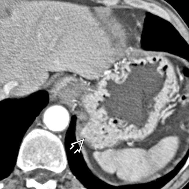
 of the stomach.
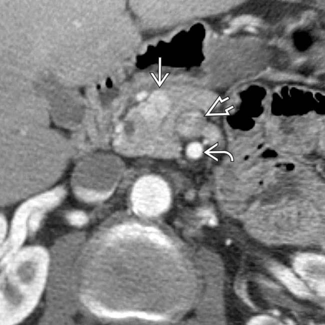
 in the pancreatic head. It is important to distinguish this from the superior mesenteric artery
  and superior mesenteric vein
  .
.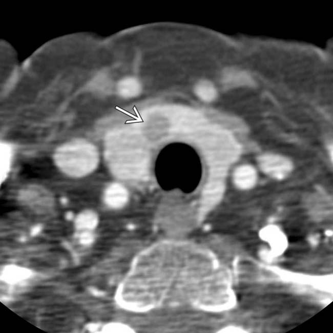
 , representing parathyroid adenomas or hyperplasia.
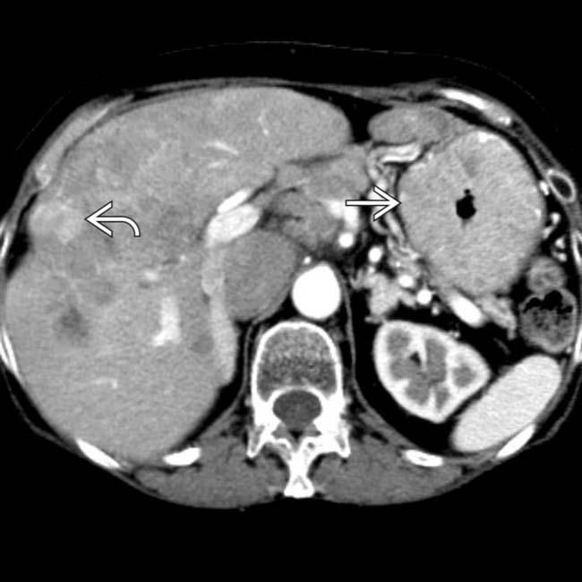
 . Multiple liver metastases are present
  . The serum gastrin levels were strikingly elevated, confirming ZES, though the gastrinoma was not identified on CT.


