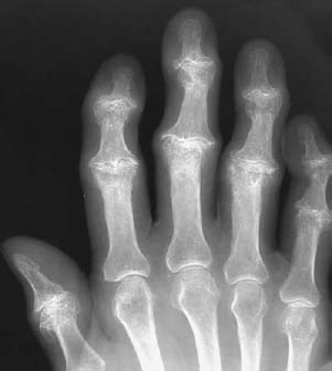
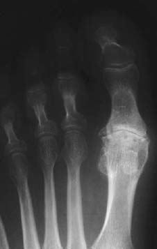
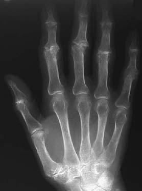
PART VII Arthritis Anthony G. Ryan and Peter L. Munk A 65-year-old man with painful hands and feet came to our institution. The request form read, “erosive arthropathy” Figure 110A Figure 110B An anteroposterior (AP) radiograph of the hand (Fig. 110A) shows marked joint space loss, articular surface irregularity, subchondral sclerosis and cyst formation, and marginal osteophyte formation at the proximal interphalangeal (PIP) and distal interphalangeal (DIP) joints of the second to fifth digits and of the interphalangeal joint of the thumb. No erosions are evident. An AP radiograph of the foot (Fig. 110B) shows the same changes to be present in the metatarsophalangeal (MTP) joint of the great toe. Osteoarthritis. Erosive osteoarthritis. Osteoarthritis is a group of conditions encompassing a primary abnormality of joints lined by hyaline cartilage and also reflecting a common final pathway of degenerative changes and attempted repair. It represents the most common arthropathy, its prevalence increasing with age. Primary osteoarthritis represents an intrinsic abnormality of articular cartilage with no superimposed associated joint disease. In this condition, spontaneous degeneration of the joint occurs without a preexisting disorder, such as trauma, and can manifest itself early in life. More commonly, secondary osteoarthritis is encountered, and it is this disease process that is most frequently encountered as individuals age or after a joint is traumatized. Obesity also significantly increases the likelihood that osteoarthritis will develop, particularly in the lower extremities. A variety of other diseases, such as acromegaly, Wilson’s disease, intra-articular hemorrhage, and chronic neurologic conditions, may contribute to the development of this process. Fundamentally, osteoarthritis represents failure of maintenance of cartilage integrity with degeneration of cartilage and reparative bone formation. In most instances, the process is noninflammatory. Typically, the disease process is slowly progressive, although severe trauma may significantly accelerate its development. Repetitive minor stress is felt to play a crucial role in the development of osteoarthritis. This presumably results in low-grade damage to the cartilage preventing normal absorption and transmission of stress through a joint. This subsequently results in trabecular fracturing as well as cartilage erosion. Cartilage itself has a limited capacity for healing. Subchondral trabeculae, however, may undergo osteoblastic repair, producing increased stiffness in the bone, thereby requiring overlying cartilage to absorb more of the stress, which in turn results in increasing damage. This downward spiral eventually leads to development of frank osteoarthritis. Patients are typically middle-aged or older. There may be a history of trauma to the joint, especially in those patients presenting at an early age. The increasing prevalence of obesity is associated with an increasing incidence of osteoarthritis, particularly in the lower extremities. A history of other predisposing conditions may be elicited, for example, prior avascular necrosis of the femoral head, acromegaly, Wilson’s disease, intra-articular hemorrhage, and chronic neurologic conditions. Erosive osteoarthritis is characterized by an abrupt onset of painful joints, with morning stiffness and occasional throbbing paresthesias of the fingertips. Failure of maintenance of cartilage integrity leads to degeneration and erosion of cartilage. Subchondral trabecular fracturing and reparative bone formation or bone resorption lead to “cyst” formation (not true cysts, as they do not have a cellular lining).
CASE 110
Osteoarthritis
Clinical Presentation


Radiologic Findings
Diagnosis
Differential Diagnosis
Discussion
Background
Etiology
Pathophysiology
Clinical Findings
Complications
Pathology
GROSS
MICROSCOPIC
Stay updated, free articles. Join our Telegram channel

Full access? Get Clinical Tree


