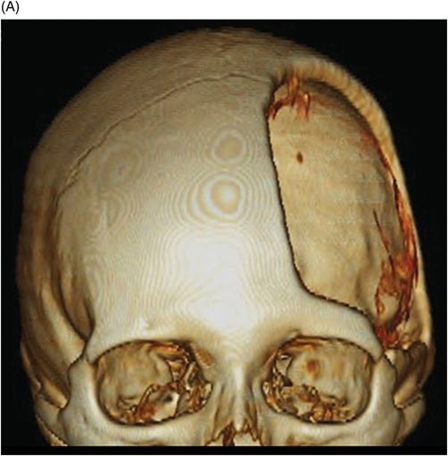
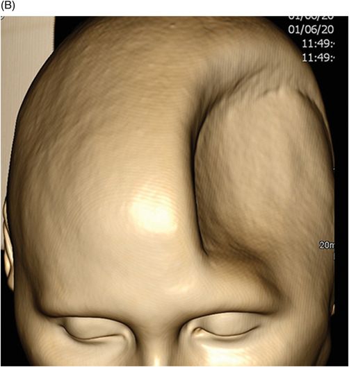
(A–B) Coronal 3D reconstruction bone and soft tissue images through the cranial vault.
Sinking Skin Flap Syndrome (Trephine Syndrome)
Primary Diagnosis
Sinking skin flap syndrome (trephine syndrome)
Differential Diagnosis
Paradoxical herniation
Imaging Findings
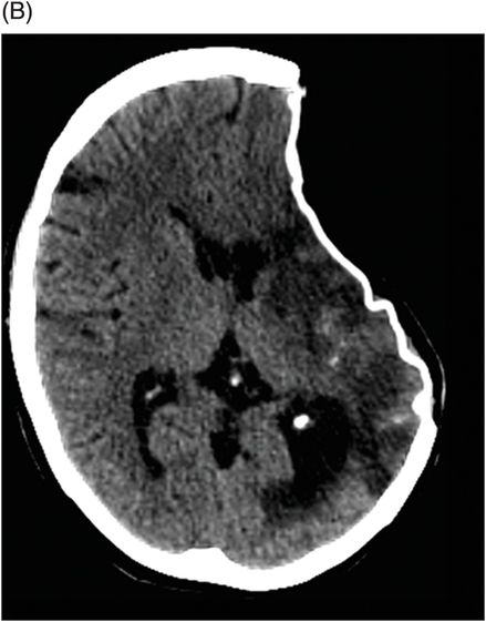
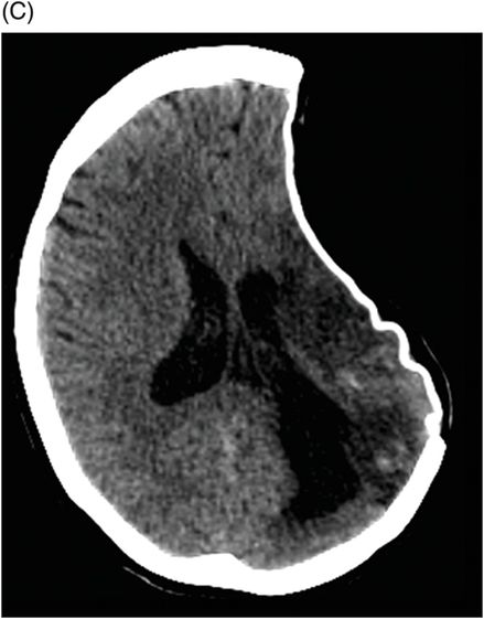
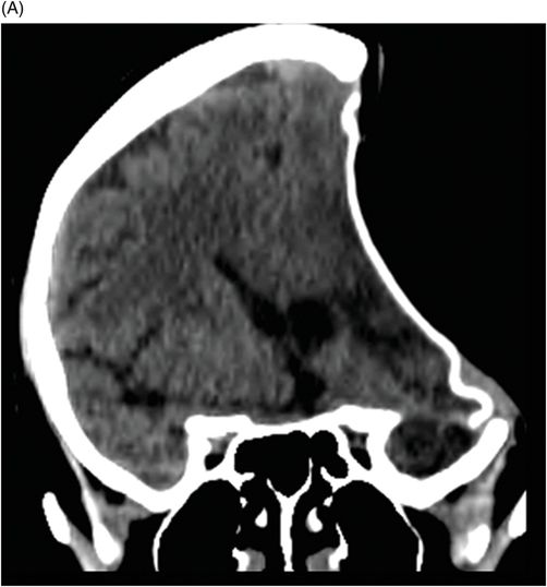
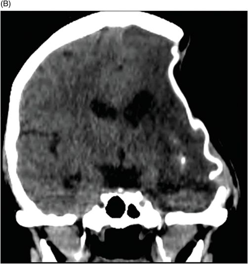
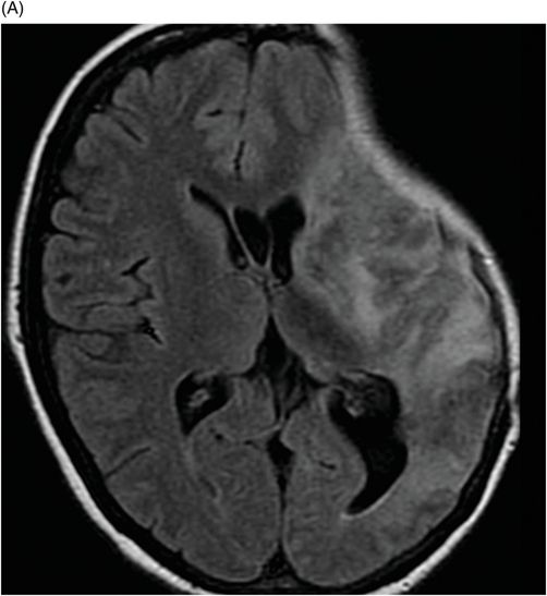
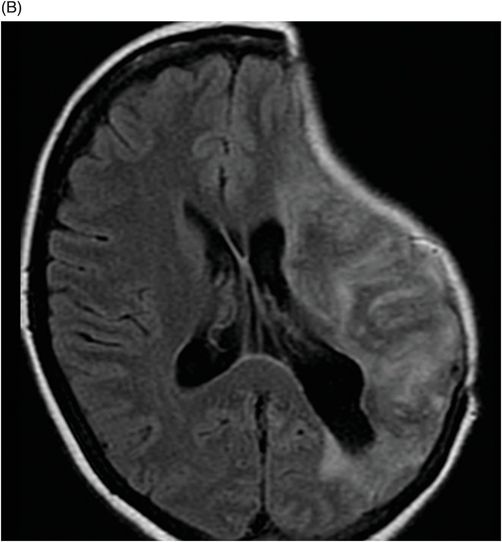
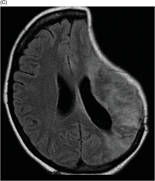
Fig. 127.1: (A–C) Axial CT images showed sinking skin flap on the left side of the cranium, characterized by the depressed meningocele complex at the craniectomy site. Fig. 127.2: (A–B) Coronal CT images confirmed the sinking skin flap on the left side of the cranium and showed concave deformity of the underlying brain parenchyma without associated signs of subfalcine or transtentorial herniations. Fig. 127.3: (A–B) 3D soft tissue and bone CT reconstruction images showed the sunken appearance of the skin flap that can be observed during physical examination. Fig. 127.4: (A–C) Axial FLAIR showed extensive cortico-subcortical hyperintensities compromising the ipsilateral temporal, frontal, parietal, and insular lobes, as well as the same side basal ganglia, corresponding to areas of gliosis/encephalomalacia, with lateral ventricle dilatation associated.


Stay updated, free articles. Join our Telegram channel

Full access? Get Clinical Tree


