Pancreas typically enlarged and edematous with loss of normal fatty lobulation
Peripancreatic fat stranding, edema, and free fluid
Mild edematous pancreatitis can appear normal on CT
• Necrotizing pancreatitis (20-30% of cases): Areas of parenchymal necrosis which are either nonenhancing or severely hypoenhancing
Differentiate cases with ≤ 30% necrosis from > 30% necrosis for patient prognosis
Necrosis may not be present initially, but can develop 3-4 days after symptom onset
• Complications
Infected pancreatic necrosis: Ectopic gas, in absence of intervention, highly suggestive of infected necrosis
Central necrosis: Necrosis of central portion of gland/duct with intact pancreas/duct in head and tail
Pseudoaneurysm: Most common locations are splenic (50%) gastroduodenal (20%), and pancreaticoduodenal (10%) arteries, but any artery can be involved
Venous thrombosis: Splenic vein most common, but portal veins or SMV can be involved
Fluid collections: Nomenclature depends on age of collection and edematous vs. necrotizing pancreatitis
PATHOLOGY
• Alcohol and gallstones account for vast majority of cases
• Many other causes, including metabolic disorders, infection, trauma, drugs, anatomic variants, neoplasm, and ERCP
CLINICAL ISSUES
• Overall mortality rate: 5%, with excellent prognosis for interstitial edematous pancreatitis
• Poor prognosis with complications: Mortality of 25% with multiorgan failure or ∼ 30% for infected necrosis
(Left) Axial CECT in an alcoholic patient demonstrates that although the pancreas itself does not appear appreciably enlarged, there is subtle peripancreatic fat stranding and edema, compatible with mild edematous pancreatitis.
(Right) Axial CECT in a patient after ERCP with placement of a stent demonstrates enlargement of the pancreas, edema with loss of normal fatty lobulation, and peripancreatic fat stranding and fluid, compatible with acute edematous pancreatitis.
(Left) Axial CECT in a patient with abdominal pain demonstrates enlargement and edema of the pancreas with surrounding fluid and stranding, compatible with acute edematous pancreatitis. The entire gland enhances normally without evidence of necrosis.
(Right) Transverse ultrasound demonstrates diffuse enlargement of the pancreas , which appears abnormally hypoechoic, compatible with acute pancreatitis in this patient with a markedly elevated lipase level.
TERMINOLOGY
Definitions
• Acute inflammation of pancreas with variable involvement of other regional tissues or remote organs
IMAGING
General Features
• Best diagnostic clue
Enlarged, edematous pancreas with peripancreatic fluid, fat stranding, and fluid collections
• Location
Pancreas and surrounding peripancreatic soft tissues
• Size
Pancreas usually increased in size (either focal or diffuse)
• Morphology
2 subtypes: Interstitial edematous, and necrotizing pancreatitis
CT Findings
• Revised Atlanta classification in 2012 standardized nomenclature used to describe acute pancreatitis
• 2 primary subtypes of acute pancreatitis
Interstitial edematous pancreatitis (70-80% of cases)
– Pancreas typically enlarged and edematous with loss of normal fatty lobulation
– Peripancreatic fat stranding, edema, and free fluid (with fluid most often localized to lesser sac, anterior pararenal spaces, and paracolic gutters)
– Usually diffuse edema of entire gland, but can rarely be focal and involve just a segment of pancreas
– Normal enhancement of pancreas without necrosis
– Normal appearance of pancreas does not exclude pancreatitis: Mild pancreatitis, usually with minimally elevated lipase levels, can appear normal on imaging
Necrotizing pancreatitis (20-30% of cases): Areas of parenchymal necrosis which are either nonenhancing or severely hypoenhancing (usually < 30 HU)
– Usually greater degree of peripancreatic fluid and inflammation than edematous pancreatitis
– Differentiate cases with ≤ 30% parenchymal necrosis from > 30% necrosis for patient prognosis
– Necrosis may not be present initially, but can develop 3-4 days after symptom onset
Early CT can underestimate or miss necrosis
– Revised Atlanta classification system describes 3 subtypes of necrotizing pancreatitis
Parenchymal necrosis alone in 5%
Parenchymal and peripancreatic necrosis in 75%
Peripancreatic necrosis alone in 20% (exudative pancreatitis)
• Complications
Infected pancreatic necrosis
– Implies superinfection of necrotic parenchyma and carries very poor prognosis
– Ectopic gas in pancreatic bed, in absence of intervention, virtually diagnostic of infected necrosis
– No other specific findings, although inflammation usually greater in cases with infected necrosis
May require aspiration for culture in cases with no definitive imaging findings
Central necrosis (disconnected duct syndrome)
– Necrosis of central portion of gland and pancreatic duct with intact upstream and downstream pancreas/duct in head and tail
– Results in fluid collection in necrotic gland with continual leakage of pancreatic juice into collection
– Diagnosis should be suggested based on distribution of necrosis
– Collection may require either internal drainage or surgery (usually distal pancreatectomy)
Extrapancreatic fat necrosis
– Due to leakage of pancreatic enzymes into peripancreatic soft tissues resulting in fat necrosis
– Usually low density with heterogeneous fluid and solid components, but can appear nodular and mass-like, mimicking carcinomatosis
– Most often occurs surrounding pancreas, anterior mesentery, or anterior pararenal spaces
– Carries better prognosis than parenchymal necrosis but worse than edematous pancreatitis
Pseudoaneurysm
– Small contrast-filled outpouching arising next to artery ± adjacent hematoma (due to leak or rupture)
– Most common locations are splenic (50%) gastroduodenal (20%), and pancreaticoduodenal (10%) arteries, but any artery can be involved
– Unexplained hemorrhage in pancreatic bed should prompt careful search for pseudoaneurysm
Venous thrombosis
– May occur due to either direct intimal injury to vessel from adjacent inflammation and pancreatic enzymes or due to mass effect from adjacent collections
– Splenic vein most often involved, but portal veins or SMV can be involved as well
Fluid collections
– Acute peripancreatic fluid collection: Fluid collection first 4 weeks after acute edematous pancreatitis
Simple, nonloculated collection of fluid attenuation with no internal debris or hemorrhage
Loculated collection with a well-defined enhancing wall of granulation tissue most often arising in lesser sac or pararenal spaces
Can rarely be found in unusual locations distant from pancreas, such as thorax
Simple collection of fluid attenuation with no internal debris or hemorrhage
– Acute postnecrotic fluid collection: Fluid collection first 4 weeks after acute necrotizing pancreatitis
Nonloculated, but containing internal necrotic debris and blood products
Acute complex fluid collection with internal debris and solid material in setting of a normally enhancing gland suggests acute postnecrotic fluid collection due to extrapancreatic necrosis
Heterogeneous collection with a well-defined wall and internal necrotic debris/blood products
– “Pancreatic abscess”: Term no longer utilized in revised Atlanta classification
“Hemorrhagic” pancreatitis:Term not included in Atlanta classification
– Small amounts of blood frequently present in peripancreatic fluid collections and has no direct impact on disease severity
MR Findings
• Pancreas appears enlarged with increased signal on T2WI and abnormally low signal on T1WI due to edema
Fat suppression very important in highlighting edema and fluid around pancreas on T2WI
• T1WI C+ images similar to CECT in detection of pancreatic necrosis and nonenhancement
• T2WI offers advantage (over CT) of allowing differentiation of simple fluid collections from collections with internal solid debris (i.e., walled off necrosis)
• MRCP can evaluate integrity of pancreatic duct, particularly in patients with suspected central gland necrosis
May be able to delineate communication between a fluid collection and pancreatic duct
Can delineate anatomic variants which might predispose to pancreatitis, including pancreatic divisum
Very sensitive for gallstones and other biliary pathology as cause of pancreatitis
• Acute pancreatitis may be associated with restricted diffusion (lower ADC values than normal pancreas)
Ultrasonographic Findings
• Enlarged, hypoechoic pancreas with adjacent free fluid and blurring of pancreatic margins
Pancreas may appear normal in mild cases
• Ultrasound often performed at presentation to look for gallstones
Radiographic Findings
• Radiography
Evidence of localized ileus due to adjacent inflammation, including dilated duodenum or sentinel loop sign (mildly dilated, gas-filled segment of small bowel ± air-fluid levels)
Colon cutoff sign: Markedly distended air-filled transverse colon with absence of gas distal to splenic flexure due to functional colonic spasm (spread of pancreatic inflammation to proximal descending colon)
Fluoroscopic Findings
• ERCP
Dilated or normal main pancreatic duct (MPD)
Communication of pseudocyst with MPD (acutely)
May show narrowed and tapered distal common bile duct (CBD) with prestenotic biliary dilatation
Angiographic Findings
• Conventional
Performed when pseudoaneurysm suspected
Useful when pancreatitis due to vascular cause
– Vasculitis, polyarteritis nodosum, lupus
– Postaortic aneurysm resection
Imaging Recommendations
• Best imaging tool
Dual-phase (arterial and venous) CECT best initial study
MR with MRCP helpful problem-solving tool to assess pancreatic duct or composition of fluid collections
DIFFERENTIAL DIAGNOSIS
Infiltrating Pancreatic Carcinoma
• Heterogeneous, hypoenhancing mass with abrupt obstruction of upstream pancreatic duct and upstream pancreatic atrophy
• Pancreatic cancer may present with pancreatitis in ∼ 5% of cases
• Focal pancreatitis can appear mass-like and mimic malignancy
• Presence of dilated pancreatic duct or biliary obstruction should prompt further investigation for underlying mass
• Pancreatic cancer infiltrates dorsally into retroperitoneum, unlike pancreatitis, which infiltrates anteriorly and laterally
• Usually other signs of malignancy, including vascular encasement, metastatic disease (most often liver), etc.


 Infected pancreatic necrosis: Ectopic gas, in absence of intervention, highly suggestive of infected necrosis
 Central necrosis: Necrosis of central portion of gland/duct with intact pancreas/duct in head and tail
 Pseudoaneurysm: Most common locations are splenic (50%) gastroduodenal (20%), and pancreaticoduodenal (10%) arteries, but any artery can be involved

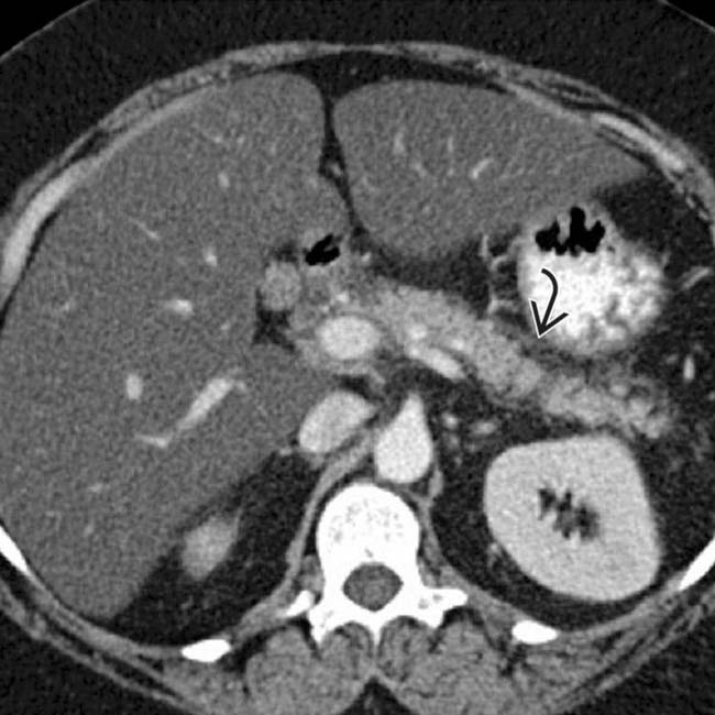
 and edema, compatible with mild edematous pancreatitis.
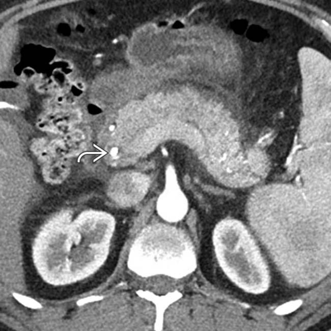
 demonstrates enlargement of the pancreas, edema with loss of normal fatty lobulation, and peripancreatic fat stranding and fluid, compatible with acute edematous pancreatitis.
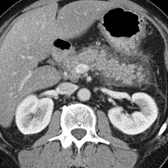
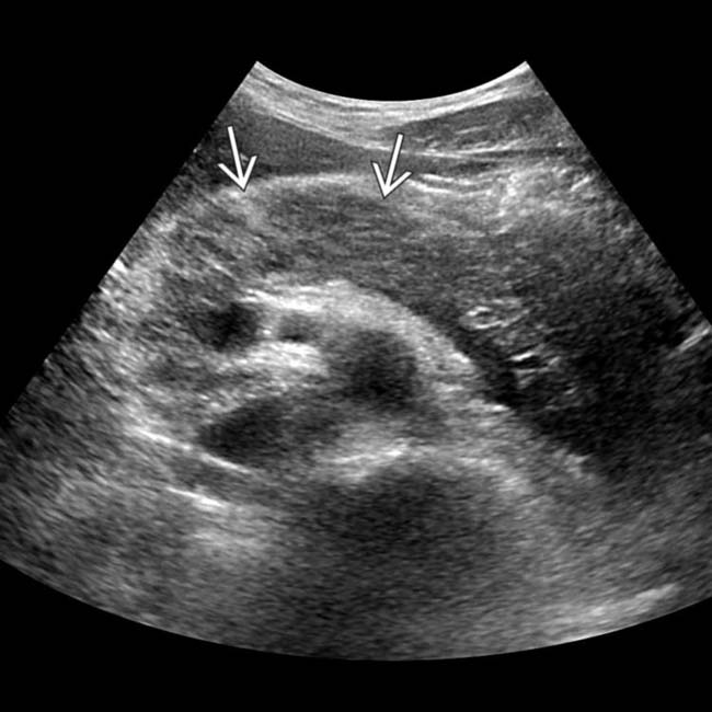
 , which appears abnormally hypoechoic, compatible with acute pancreatitis in this patient with a markedly elevated lipase level.


 Interstitial edematous pancreatitis (70-80% of cases)


 Infected pancreatic necrosis
 Central necrosis (disconnected duct syndrome)
 Extrapancreatic fat necrosis
 Pseudoaneurysm
 Venous thrombosis
 Fluid collections

 Loculated collection with a well-defined enhancing wall of granulation tissue most often arising in lesser sac or pararenal spaces


 Can delineate anatomic variants which might predispose to pancreatitis, including pancreatic divisum


