• MR is a good alternative to CT in pregnant patients, and children when US is nondiagnostic
TOP DIFFERENTIAL DIAGNOSES
• Mesenteric adenitis and enteritis
• Ileocolitis
• Crohn disease
• Gynecologic causes
• Cecal diverticulitis
• Appendiceal tumor
• Cecal carcinoma
• Cystic fibrosis
CLINICAL ISSUES
• Clinical diagnosis is incorrect in ∼ 20% of young men and 40% of young women
• Also frequently in error in young children and older adults
(Left) This graphic illustrates some of the characteristic features of acute appendicitis, including the distended, thick-walled, inflamed appendix and inflammatory thickening of the adjacent walls of the cecum and terminal ileum .
(Right) Axial CECT in 12-year-old boy shows perforated appendicitis. Note the appendicolith and cecal wall thickening . The focally necrotic wall of the appendix delineates the point of perforation. Pericecal and periappendiceal inflammatory changes are also evident.
(Left) Longitudinal sonogram demonstrates a distended, thick-walled appendix , 10 mm in diameter with adjacent hyperechoic periappendiceal inflammation of fat , indicative of an inflammatory process and diagnostic for appendicitis.
(Right) Color power Doppler sonography in a patient with acute appendicitis demonstrates marked hyperemia in the wall of the appendix , indicative of inflammation and consistent with acute appendicitis.
TERMINOLOGY
Definitions
• Acute appendiceal inflammation due to luminal obstruction and superimposed infection
IMAGING
General Features
• Best diagnostic clue
Distended, thick-walled, noncompressible appendix (≥ 7 mm) on US
Abnormal mural enhancement of distended appendix on CECT
Periappendiceal fat stranding on US or CT
• Location
Appendix arises from cecal tip, but tip may lie some distance from cecum
• Size
Noncompressible appendix on US
– > 6 mm has sensitivity of 100%, but specificity of only 64%
– > 7 mm has sensitivity of 94% and specificity of 88%
– 6-7 mm equivocal size; increased flow on color Doppler in appendix indicates positive study
• Morphology
Tip of appendix often 1st site of inflammation and appendiceal perforation
Radiographic Findings
• Radiography
Appendicolith in < 5% of patients (on plain films)
Air-fluid levels within bowel in RLQ
– Due to focal ileus
Loss of right psoas margin
Appendiceal lumen may be patent to point of obstruction
With perforation
– Small bowel obstruction (SBO)
– RLQ extraluminal gas (uncommon to rare)
– Displacement of bowel loops from RLQ
CT Findings
• NECT
Dilated appendix ≥ 7 mm
Periappendiceal fat stranding
Appendicolith
– Seen much more frequently on CT than on radiography
– Even noncalcified appendicoliths may be seen on CT
With perforation
– Small bowel obstruction
– Inflammatory phlegmonous or fluid collections demonstrating mass effect, most commonly in RLQ or dependent pelvis (cul-de-sac)
• CECT
Dilated appendix ≥ 7 mm
– Sensitivity 95%, specificity 95%
Abnormal enhancement of appendiceal wall on CECT
– Inflamed mucosa may show hyperenhancement
– Necrotic wall may show no enhancement
– Wall may be discontinuous at point of perforation
± bubbles of extraluminal gas
Appendicolith may be present (15-40%)
Focal bowel wall thickening of adjacent wall of cecum &/or terminal ileum
– Focal ileus of distal small bowel due to inflammation and spasm of lumen
– Frank SB obstruction may occur with abscess or delayed diagnosis
RLQ extraluminal inflammation
– Diffuse nonencapsulated inflammation of mesoappendix, mesenteric and omental fat (“phlegmon”)


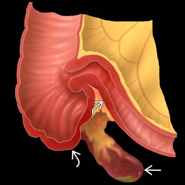
 and inflammatory thickening of the adjacent walls of the cecum and terminal ileum
  .
.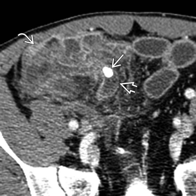
 and cecal wall thickening
  . The focally necrotic wall of the appendix
  delineates the point of perforation. Pericecal and periappendiceal inflammatory changes are also evident.
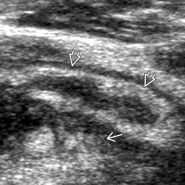
 , 10 mm in diameter with adjacent hyperechoic periappendiceal inflammation of fat
  , indicative of an inflammatory process and diagnostic for appendicitis.
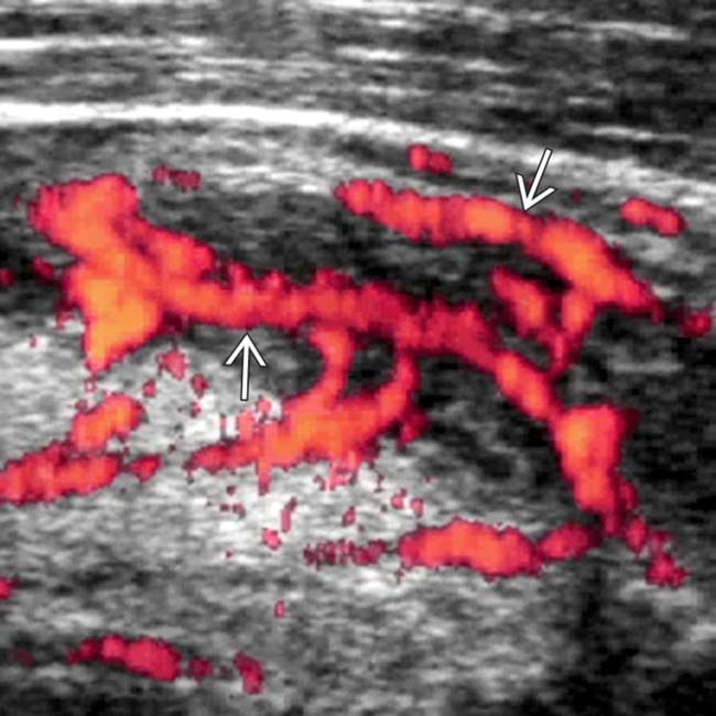
 , indicative of inflammation and consistent with acute appendicitis.


 Noncompressible appendix ≥ 7 mm


