Biological Effects of Magnetic Resonance Imaging
Objectives
At the completion of this chapter, the student should be able to do the following:
Key Terms
Many noninvasive medical imaging modalities are currently available, such as x-ray imaging, radioisotope imaging, ultrasonography, and magnetic resonance imaging (MRI). Being noninvasive, each of these modalities is inherently safe. Ionizing radiation can induce malignant disease and genetic mutations, although the risk of such responses is extremely low.
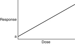
Human response to ionizing radiation follows a linear, nonthreshold, dose-response relationship (Figure 29-1). Nonthreshold means that no dose of ionizing radiation is considered to be absolutely safe. Even the smallest dose carries a risk of response, although such risk may be lower than that from other everyday risks.
At low radiation doses, the increase in response is exceedingly low. Only after rather high radiation doses (e.g., 0.25 Gy or greater) would the anticipated response be detectable.
Such responses to ionizing radiation are stochastic. There is no threshold dose, and the response is an increase in incidence, such as cancer or leukemia, rather than severity.
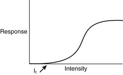
Exposure to the energy fields of MRI, both individually and collectively, results in a fundamentally different type of dose-response relationship (Figure 29-2). Such a dose-response relationship is known as a threshold, nonlinear relationship.
It is inappropriate to use the term dose when describing the energy fields associated with MRI. Intensity is a better term, because it implies bathing a patient in an agent, rather than administering a certain quantity of the agent to the patient.
Regardless of the type of MRI energy field considered, there is a level of intensity, the threshold intensity (IT), below which no response is elicited. Below IT, MRI is entirely safe. Above IT, the response to MRI exposure first increases slowly and then more rapidly until 100% response is observed. A response that exhibits a threshold and a dose-related severity is a deterministic response.
Because MRI is still developing, all of the radiobiological questions are not completely answered. It does appear that MRI, as it is currently used, is safe for all workers and patients. Experimental observations suggest that IT for all of the MRI energy fields is considerably higher than the intensities used for clinical examinations.
Magnetic Resonance Imaging Energy Fields
Although MRI is considered safe, radiobiologists will be busy for many years working to stretch their ability to detect human responses to the energy fields used. It is certain that the delayed determination that x-ray exposure can cause cancer and leukemia will not be repeated with MRI, because the energy fields are nonionizing.
The potential health hazards of MRI lie in the static magnetic field (B0), the transient gradient magnetic fields (BSS, BΦ, BR), the radiofrequency (RF)-pulsed electromagnetic field, or a combination of these. Radiobiological data already show that, individually, such fields are innocuous at the levels currently used for MRI.
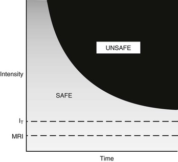
In addition to following a threshold intensity-response relationship, these fields exhibit a time-intensity relationship (Figure 29-3). The IT applies to continuous exposure. Above this threshold, shorter exposure times produce the same response. What is not fully understood is whether these MRI energy fields have adverse health effects when combined.
Before there is a discussion of suspected biological effects of MRI in human beings, an examination of each of the MRI fields and their potential effects on other biological systems is necessary. The three MRI energy fields interact with matter differently (Table 29-1).
TABLE 29-1
Principal Mechanisms of Interaction of the Three Magnetic Resonance Imaging Energy Fields with Tissue
| Magnetic Resonance Imaging Energy Field | Mechanism of Interaction |
| Static magnetic field (B0) | Polarization |
| Transient gradient magnetic fields (BSS, BΦ, BR) | Induced currents |
| RF field (RFt) | Thermal heating |
Static Magnetic Fields
The static magnetic field is probably the best-recognized MRI energy field. Although people cannot sense a magnetic field, they know that it exists because of forces that attract and repel small magnets, such as kitchen latches and other everyday permanent magnets. Consequently, it is also clear that one interaction between a static magnetic field and matter results in a force that can turn certain types of matter (ferromagnetic material) into projectiles or missiles.
Additionally, tissue can be rendered weakly magnetic, or polarized, by exposure to a strong static magnetic, field (B0). This induced tissue magnetism cannot be sensed. Furthermore, in the presence of a static magnetic field, tissue magnetization (M) can be measured only with great difficulty in MRI. Once removed from the influence of a static magnetic field, tissue relaxes to its normal state without a hint that it was previously magnetized.
There is no scientific evidence to show that intense static magnetic fields produce harmful effects in mammals. Mechanisms have been suggested and subjected to considerable investigation but the result remains negative.
There are few studies of the effects of long-term exposure of mammals to intense static magnetic fields that use proper controls and large sample sizes. Some experiments have been designed to detect growth abnormalities, biochemical disruption, and malignant disease induction in rodents at magnetic field strengths up to 5 T. All such observations have been negative.
Other reports deal with the effects of intense static magnetic fields on cultured cells. These reports do not suggest any adverse effects on cell growth for magnetic fields up to 7 T. Such experiments involve conventional culturing of human fibroblasts. Lengthening the exposure time does not produce a positive response either.
The navigational ability of adult bees and pigeons is considered to be mediated by granules of magnetite deposits in the abdominal region and in the head, respectively (Figure 29-4). These magnetic deposits serve as a built-in compass, yet experiments have not been conducted to determine whether these characteristics can be disturbed by intense static magnetic fields. Such experiments are not pertinent anyway because human beings do not rely on an internal compass.
Some speculation exists concerning bioeffects of intense magnetic fields on growth rate, mutation, fertility, and blood cell count in mammals. Growth rate and mutation effects have been reported in animals and yeast, but the results have been equivocal or not reproducible. Monkeys who were exposed to a static magnetic field of 7 T for up to 1 hour experienced a transient decrease in heart rate and sinus arrhythmia; a later study failed to demonstrate these effects with a limited exposure of 15 minutes at 10 T.
The evidence for static magnetic field effects on the developing embryos of pregnant mice who were exposed at various times during gestation did not consistently show alterations in the offspring when compared with nonexposed control mice. These studies used acceptably large sample sizes and were repeated with similar negative results.
Transient Magnetic Fields
The gradient magnetic fields used to spatially encode the magnetic resonance (MR) signals vary with time. These are often termed moving magnetic fields or time-varying magnetic fields. The preferred term is transient magnetic field.
A transient magnetic field is able to induce an electric current. For example, an electric current can easily be induced in a metal conductor. This is the basic principle of electromagnetic induction that generates and detects the MR signal.
Theoretically, transient magnetic fields can either stimulate or impair electric impulses along neuronal pathways in tissue. The main influence of this is the time rate of change of the magnetic field expressed as dB/dt and measured in T/s. For example, a transient magnetic field of 3 T/s results in an induced electric current density of approximately 3 µA/cm2 in tissue.
A current density of 3 µA/cm2 is capable of producing involuntary muscle contraction and cardiac fibrillation in experimental animals. However, the time of application was far greater than that experienced in clinical MRI.
Because some cells and tissues are electric conductors, the transient gradient magnetic fields may induce or interfere with normal conduction pathways. Depending on the intensity of the induced electric current, the normal function of nerve cells and muscle fibers may be affected. The threshold current density of transient magnetic fields depends on the tissue conductivity and the duration of time the field is energized.
The maximum transient magnetic field does not occur in the center of the magnet. There the gradient magnetic fields are near zero; they are maximum at the periphery of the imaging volume.
Radiofrequency Fields
The radiofrequency field (RF) used in MRI exists in the approximate range of 10 to 200 MHz. This energy field consists of an oscillating electric field and a similar orthogonal oscillating magnetic field. These fields interact simultaneously with matter. The interaction manifests differently according to the nature of matter.
The electric field component can induce heating in matter and induce high-frequency electric currents, whereas the magnetic field component acts as a rapidly transient magnetic field. This is relevant to MRI because it has the potential for heating both superficial and deep tissues.
The electric field is generally well shielded from the patient’s body by the placement of guard rings in the RF coil design. The magnetic component of the RF field is required for imaging. However, in addition to flipping proton spins, it induces eddy currents within the conductive tissues of the body. This results in heat, which increases with increasing frequency.
A rise in body temperature has been reported with exposure to RF fields; the absorbed energy causes this temperature increase. In turn, the RF frequency, exposure time, and mass of the exposed object determine the amount of absorbed energy.
The RF frequency influences energy absorption because it is wavelength dependent. Energy is most efficiently absorbed from an RF emission when the tissue size is approximately one half of the RF wavelength. The wavelengths associated with 10 MHz and 200 MHz are 30 m and 1.5 m, respectively. Shorter wavelength RF results in more superficial heating and less penetration.
Commercial microwave ovens operate at 2.45 GHz. What is the wavelength of such radiation? … 12 cm. What is the size of a hot dog? … 12 cm.
Depending on the imaged region, when an RF pulse sequence is applied to a patient the peak power can range to 16 kW. The energy returned from the patient is very weak; it is measured in microwatts (µW). Some energy is lost to coil inefficiencies and cable losses; some is reflected back to the RF power amplifier and a load attached to the back of the coil.
Some energy is transmitted through the patient without interactions, and some energy interacts with the patient and generates heat, causing a possible rise in body temperature. The amount of absorbed energy depends on the transmitted magnetic field frequency, the total power of the RF pulse, and the structure and conductivity of the exposed object.
The SAR has units of W/kg and may be expressed as an average over the whole body or any specific tissue. Thus there is a whole-body and a local SAR.
The SAR may also be expressed over a long time or a single pulse. Specific absorption rate is a measure of the energy absorbed per unit time per unit mass.
Specific absorption rate is to MRI as the Gray is to ionizing radiation. The mathematical formulation of SAR is extensive and unnecessary for this discussion.
Biological effects of RF are associated with the SAR. In turn, the SAR is related to the RF power density because it varies in time (temporal) and space (spatial). Recommended exposure limits are expressed as power density. They are set approximately 100 times lower than levels known to cause a biological response.
Stay updated, free articles. Join our Telegram channel

Full access? Get Clinical Tree


