• Intrahepatic and systemic venous collaterals bypass obstructed hepatic veins and IVC
Spider web pattern of hepatic venous collaterals on CT, MR, angiography
• Large regenerative nodules (form of nodular regenerative hyperplasia) are characteristic of chronic BCS
Imaging and histology similar to FNH
May have peripheral halo and central scar
Hypervascularity persists into venous phase without washout
Uniform or peripheral delayed retention (bright) on gadoxetate-enhanced MR
• Absent, reversed, or flat flow in hepatic veins; reversed flow in IVC on color Doppler US
PATHOLOGY
• Etiology in western populations is usually a hypercoagulable condition
DIAGNOSTIC CHECKLIST
• Do not mistake BCS for cirrhosis
Pathogenesis, imaging findings, prognosis, and treatment are very different
• Do not mistake caudate hypertrophy or large regenerative nodules for hepatocellular carcinoma
• Check for hypercoagulable conditions, prior chemotherapy, or bone marrow transplant
(Left) Axial anatomic illustration of Budd-Chiari syndrome demonstrates ascites, venous collaterals , heterogeneous hepatic parenchyma due to centrilobular necrosis, and hypervascular regenerative nodules . Note the sparing of the caudate lobe with hypertrophy , as well as the thrombosed IVC.
(Right) Axial CECT shows caudate hypertrophy, a large caudate collateral vein , and peripheral atrophy and heterogeneity. The hepatic veins were occluded.
(Left) Transverse color Doppler ultrasound of the liver in a 48-year-old woman with known polycythemia vera, RUQ pain, and elevated liver function tests reveals a lack of flow within the right hepatic vein .
(Right) Color Doppler ultrasound in the same patient demonstrates a large intrahepatic collateral vein bypassing the occluded hepatic veins.
TERMINOLOGY
Abbreviations
• Budd-Chiari syndrome (BCS)
Synonyms
• Hepatic venous outflow obstruction
Definitions
• Global or segmental hepatic venous outflow obstruction
At level of large hepatic veins or suprahepatic segment of inferior vena cava (IVC)


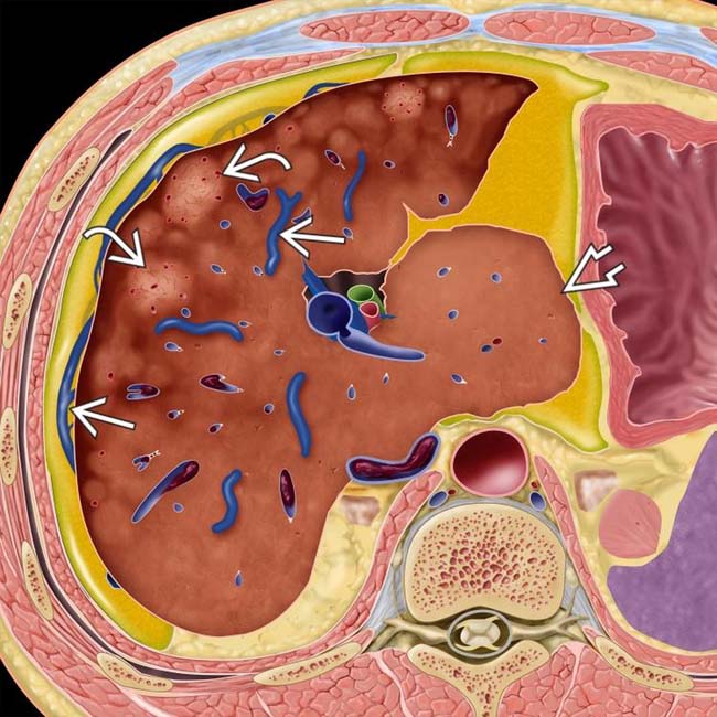
 , heterogeneous hepatic parenchyma due to centrilobular necrosis, and hypervascular regenerative nodules
  . Note the sparing of the caudate lobe with hypertrophy
  , as well as the thrombosed IVC.
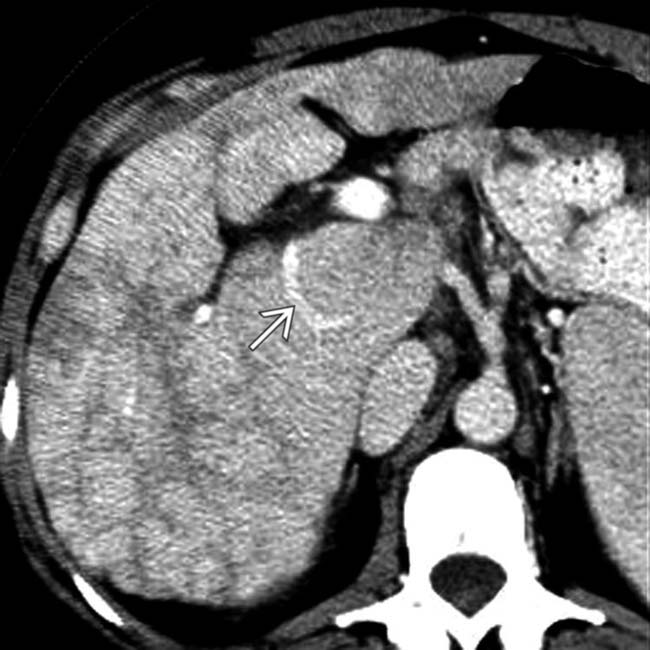
 , and peripheral atrophy and heterogeneity. The hepatic veins were occluded.
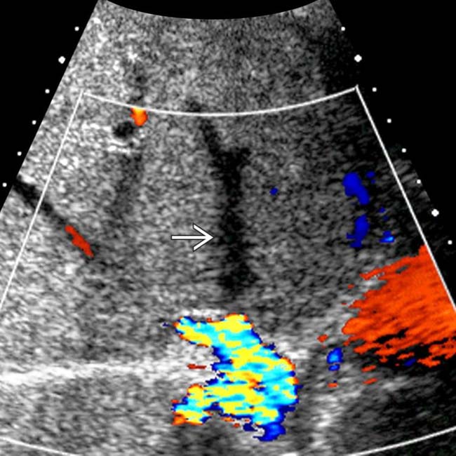
 .
.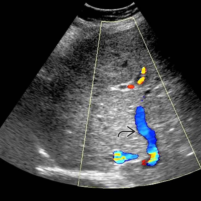
 bypassing the occluded hepatic veins.


 Long segmental compression or stenosis of IVC


