Contrast Agents and Magnetic Resonance Imaging
Objectives
At the completion of this chapter, the student should be able to do the following:
• List two general approaches to magnetic resonance imaging (MRI) contrast enhancement.
• Identify three routes for administration of MRI contrast agents and give an example of each.
• Describe paramagnetism and how such an agent enhances image contrast.
• Identify the blood–brain barrier and how it affects the use of MRI contrast agents.
Key Terms
With magnetic resonance imaging (MRI), the naturally available contrast among tissues in the body is good. It was originally assumed that contrast enhancement (CE) would not be needed in MRI. Even without the use of a contrast agent, MRI is exceptionally sensitive to detecting pathologic conditions. However, as the field develops, it is becoming clear that under some circumstances the addition of a contrast agent greatly increases the diagnostic value of MRI by improving disease sensitivity and specificity and by delineating pathologic processes.
In some areas of the body, adjacent tissues have similar MRI appearance, and CE is needed for adequate differentiation. CE is also useful for delineating the structural integrity of the blood–brain barrier. The addition of a contrast agent allows imaging of areas of normal versus decreased tissue perfusion. In some cases, pathology can be better identified because of the different speeds with which a contrast agent enters or washes out of tumors as compared with the surrounding normal tissue.
The properties needed in an MRI contrast agent are the same as those needed for contrast agents in other imaging modalities. The agent should alter contrast at low concentrations, and its effect on contrast should be dose dependent. It must be nontoxic and nonreactive, as well as rapidly cleared from the body.
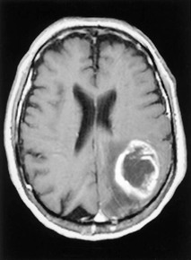
The biodistribution of a contrast agent is exceedingly important. An ideal contrast agent distributes only to the organ, tissue, or pathology of interest (Figure 27-1).
Approches to Contrast Enhancement
The normal MRI appearance of a tissue depends on both the physical characteristics of the tissue (such as viscosity and temperature) and the chemical characteristics that influence proton density (PD), spin-lattice relaxation (T1), and spin-spin relaxation (T2).
Although changing a physical property of a sample is useful for in vitro studies, it is generally not safe to use a sufficiently large change in a living organism to make this approach useful in vivo. However, it is possible to safely alter the chemical characteristics of living tissue sufficiently to change the MRI parameters and therefore image appearance.
In MRI, the image contrast is influenced by a number of factors, including the hydrogen content (PD), spin-lattice relaxation (T1), spin-spin relaxation (T2), flow through the area being examined, and the magnetic susceptibility of the tissue. If any of these parameters are changed by the addition of a contrast agent, the magnetic resonance (MR) image appearance is also changed.
When the use of an MRI contrast agent causes the tissue of interest to appear brighter, positive CE occurs. The CE is negative if the tissue of interest is darker when a contrast agent is used.
Although MRI contrast agents have traditionally been used for positive contrast, it is becoming more common to use them to reduce contrast, as in susceptibility-weighted cerebral perfusion imaging. Box 27-1 presents one classification for MRI contrast agents.
Altering Hydrogen Content
The most obvious approach to CE is to alter the hydrogen content (PD) of tissue. Examples of this approach are water loading to increase the total signal from the kidneys and bladder and administering diuretics to decrease hydration of tissue.
An alternative approach is to remove the source of the signal entirely by administering a hydrogen-free contrast agent that fills the area of interest. Another approach is to use oxygen. Unfortunately, although oxygen is paramagnetic, the effect is too weak for clinical use.
The amount of MR signal arising from tissue can also be changed by altering the ratio of water to fat in that tissue. The relaxation characteristics of fat and water are quite different, so greatly increasing the amount of fat can increase the available signal. This approach has been used for imaging of the gut, with oral administration of fat-containing compounds such as mineral oil.
Altering the Local Magnetic Field
Motion Reduction.
One approach to changing the magnetic environment perceived by spins is to administer an agent that greatly slows the normal motion of hydrogen. Normally, a hydrogen ion is tumbling quickly enough that any variations in the local magnetic environment are averaged (rather like the blurring in vision that a person experiences when spinning around rapidly).
Stay updated, free articles. Join our Telegram channel

Full access? Get Clinical Tree


