Echo-Planar Imaging
Objectives
At the completion of this chapter, the student should be able to do the following:
Key Terms
Echo-planar imaging (EPI) is a method for extremely fast formation of the magnetic resonance (MR) image. Some of its commercial implementations have trade names that imply instantaneous image acquisition. Although that may be a slight exaggeration, imaging times of as little as 50 ms to produce images of moderate quality are realistic.
Echo-Planar Imaging
In 1977 Mansfield first introduced the concept of EPI. The distinguishing characteristic of EPI is the filling of k-space after a single radiofrequency (RF) excitation. Each line of k-space does not require separate spin excitation as in spin echo (SE) or gradient echo (GRE) imaging.
The entirety of k-space can be sampled and filled by measuring either the envelope of gradient-refocused free induction decay (FID) or that of an SE. In either case, the entire image is acquired following a single RF excitation and within one repetition time (TR).
 The fundamental idea of EPI is to fill k-space with the magnetization produced by a single RF pulse.
SE and GRE techniques measure only a portion of k-space in each TR interval. That portion is typically one Fourier line of k-space. Thus T1 is a consideration because it is necessary to allow longitudinal magnetization to recover before the next portion of k-space can be measured.
EPI avoids this problem by measuring all of k-space in one pass. However, because this one pass may be rather long, spin-spin relaxation (T2) during signal sampling becomes an issue. The raw data measurements must be made rapidly to measure all of k-space before the transverse magnetization MXY is substantially altered in magnitude by T2* relaxation.
Thus the magnetic resonance imaging (MRI) system must be capable of extremely fast and high-amplitude gradient switching and rapid data acquisition. Manufacturers have solved these engineering and hardware requirements.
A single RF excitation α pulse is followed by a 180° RF refocusing pulse. The MR signal read phase contains a train of GREs produced by rapidly switching the read gradient magnetic field (GR) (Figure 20-1).
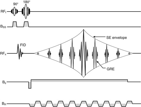
After a single excitation, the read gradient is switched from one polarity to another, generating a GRE each time. In its original form, demonstrated by Mansfield, the phase encoding is done with a small constant gradient throughout the acquisition. This way, each echo has a different phase encoding and can be used to fill k-space. Acquisition times are about 10 times faster than those for the GRE imaging techniques described in Chapter 18.
The train of echoes obtained in EPI is produced by switching the gradient magnetic field and sampling the signal in the envelope of the SE. EPI GREs are produced with modifications of the scheme shown in Figure 20-1.
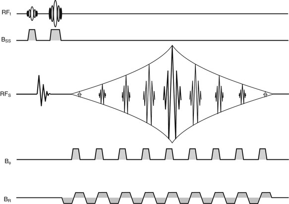
The original echo-planar method proposed by Mansfield was complicated by limitations of his hardware. A more recent version, blipped echo planar, has been universally adopted in commercial MRI systems.
Figure 20-2 shows the pulse sequence diagram of a blipped echo-planar image. Figure 20-3 shows the path through k-space that is traversed in sampling the GRE signals.
Related posts:

Stay updated, free articles. Join our Telegram channel

Full access? Get Clinical Tree


