• MR shows some elements better than CT (lipid and hemorrhage)
• Gadoxetate-enhanced MR (Eovist; Primovist)
Adenoma shows no substantial uptake or retention
Key distinction from FNH
• T1WI: Mass: Heterogeneous signal intensity
Increased signal intensity (due to fat or recent hemorrhage)
Decreased signal intensity (necrosis, calcification, old hemorrhage)
• Heterogeneous, hypervascular mass with foci of fat or hemorrhage in a young woman
TOP DIFFERENTIAL DIAGNOSES
• Hepatocellular carcinoma (HCC)
HCC typically occurs in older, cirrhotic men
• Fibrolamellar HCC
• Focal nodular hyperplasia
Homogeneously enhances; retains gadoxetate
• Hypervascular metastases
PATHOLOGY
• Hepatic steatosis, pregnancy, anabolic steroids, and oral contraceptives increase number and growth rate of adenomas
CLINICAL ISSUES
• Risk factors for HCC
Large adenoma, male sex, glycogen storage disease, anabolic steroid use, CTNNB1 -mutated subtype of HA
(Left) Graphic shows a hypervascular mass in the right lobe and spontaneous subcapsular bleeding .
(Right) Axial CECT of a 40-year-old woman with sudden RUQ pain and syncope shows an intensely enhancing mass in the right lobe of the liver. A lentiform heterogeneous collection of fluid indents the surface of the liver, and within this collection is a focus of higher density likely representing a sentinel clot. A ruptured inflammatory hepatic adenoma was resected.
(Left) Photograph of a resected specimen shows a large adenoma with central areas of rupture and hemorrhage . (Courtesy M. Yeh, MD, PhD.)
(Right) Photomicrograph of a hepatic adenoma features a thin-walled unpaired vessel surrounded by neoplastic hepatocytes with abundant steatosis. Imaging often reveals these features, directly or indirectly. (Courtesy M. Yeh, MD, PhD.)
TERMINOLOGY
Abbreviations
• Hepatic adenoma (HA)
Synonyms
• Hepatocellular adenoma, liver cell adenoma
Definitions
• Heterogeneous group of benign hepatocellular neoplasms with distinctive genetic, pathologic, and clinical features
IMAGING
General Features
• Best diagnostic clue
Heterogeneous, hypervascular mass with foci of fat or hemorrhage in a young woman
• Location
Subcapsular region of right lobe of liver (75%)
Intraparenchymal or pedunculated (10%)
• Size
Varies from 6-30 cm
• Key concepts
Very uncommon relative to focal nodular hyperplasia (FNH) and hepatocellular carcinoma (HCC)
3 distinct subtypes with different genetics, pathology, clinical features
CT Findings
• Depending on HA subtype
Encapsulation seen in ∼ 20%, best on delayed phase CECT
Hemorrhage within tumor, best seen on NECT as hyperdense foci
Intratumoral lipid, best seen on NECT as hypodense foci
Hypervascularity
– Most intense and persistent in inflammatory subtype of HA
Calcification: Focal, present in ∼ 5%
MR Findings
• T1WI
Mass: Heterogeneous signal intensity
– Increased signal intensity (due to fat and recent hemorrhage), more evident on MR than CT
– Decreased signal intensity (necrosis, calcification, old hemorrhage)
Rim (fibrous pseudocapsule): Hypointense
• T2WI
Mass: Heterogeneous signal intensity
– Increased signal intensity (old hemorrhage, necrosis)
– Decreased signal intensity (fat, recent hemorrhage)
Rim (fibrous pseudocapsule): Hypointense
• T1WI C+
Gadolinium, arterial phase
– Heterogeneous hypervascular enhancement (especially in inflammatory subtype)
Delayed phase
– Pseudocapsule: Hyperintense to liver and adenoma
• Gadoxetate-enhanced MR (Eovist, Primovist)
Hepatocellular-specific contrast agent
Adenoma shows no substantial uptake or retention on delayed imaging
– Key distinction from FNH
Ultrasonographic Findings
• Grayscale ultrasound
Complex, hyper-/hypoechoic, heterogeneous mass with anechoic areas
– Due to fat, hemorrhage, necrosis, and calcification
– Capsule may be seen
• Color Doppler
Hypervascular tumor
Large peripheral arteries and veins
Intratumoral veins present
– Absent in FNH; useful distinction for adenoma
Angiographic Findings
• Conventional
Hypervascular mass with centripetal flow
Enlarged hepatic artery with feeders at tumor periphery (50%)
Hypovascular; avascular regions
– Due to hemorrhage and necrosis
Nuclear Medicine Findings
• Technetium sulfur colloid (TcSC)
Usually “cold” (photopenic) (80%)
Uncommonly “warm” (20%)
– Due to uptake in sparse Kupffer cells
• HIDA scan
Increased activity in some
• Gallium scan
No uptake
Imaging Recommendations
• Best imaging tool
Gadoxetate-enhanced MR, including multiphasic and delayed imaging
In- and opposed-phase GRE
DIFFERENTIAL DIAGNOSIS
Hepatocellular Carcinoma
• May be hard to distinguish on imaging or pathology
• Biliary, vascular, nodal invasion and metastases = malignancy
• HCC typically occurs in older, cirrhotic men
Adenoma occurs in young, healthy women
Fibrolamellar HCC
• Large, lobulated mass with scar and septa
• Vascular, biliary invasion and metastases common
Focal Nodular Hyperplasia
• Arterial phase: Homogeneously enhancing mass
Only gold members can continue reading. Log In or Register to continue


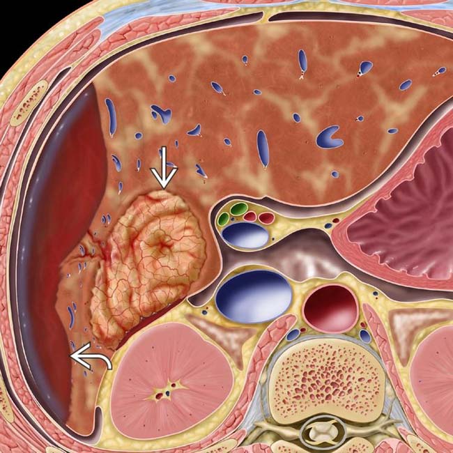
 in the right lobe and spontaneous subcapsular bleeding
  .
.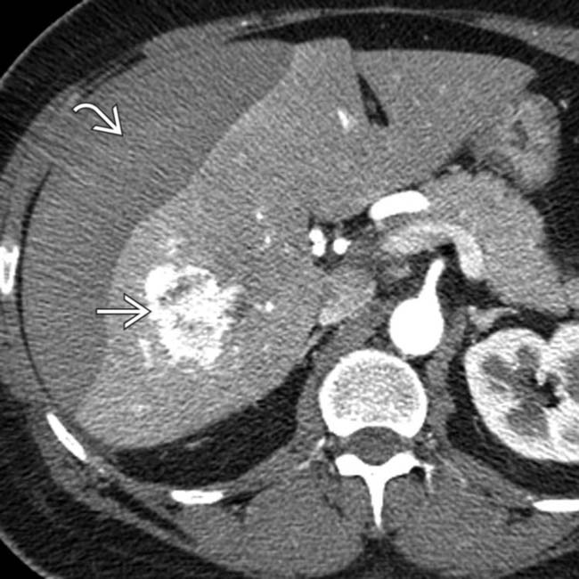
 in the right lobe of the liver. A lentiform heterogeneous collection of fluid indents the surface of the liver, and within this collection is a focus of higher density
  likely representing a sentinel clot. A ruptured inflammatory hepatic adenoma was resected.
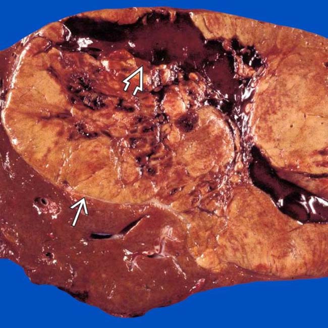
 with central areas of rupture and hemorrhage
  . (Courtesy M. Yeh, MD, PhD.)
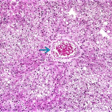
 surrounded by neoplastic hepatocytes with abundant steatosis. Imaging often reveals these features, directly or indirectly. (Courtesy M. Yeh, MD, PhD.)


