Arterial phase: Early peripheral, nodular or globular, discontinuous enhancement
• Small hemangiomas (capillary): < 2 cm
Arterial and venous phases: Homogeneous enhancement (flash-filling)
• Typical hemangiomas: 2-10 cm in diameter
Venous phase: Progressive centripetal enhancement to uniform filling, still isodense to blood vessels
• Giant hemangioma: > 10 cm in diameter
Venous and delayed phases: Incomplete centripetal filling of lesion (scar does not enhance)
• US: Peripheral rim or homogeneously hyperechoic mass ± acoustic enhancement
TOP DIFFERENTIAL DIAGNOSES
• Cholangiocarcinoma (peripheral)
• Hypervascular metastases
• Hepatic angiosarcoma
DIAGNOSTIC CHECKLIST
• Small hepatocellular carcinomas and hypervascular metastases
Can mimic small hemangiomas by their uniform homogeneous enhancement pattern
• Hemangiomas
Remain isodense to blood vessels on portal venous and delayed phases of enhancement
• Other benign and malignant liver masses
Usually become hypodense to blood vessels and liver (except cholangiocarcinoma)
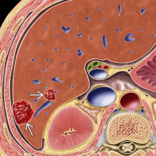
(Left) Graphic shows 2 hemangiomas as nonencapsulated collections of blood within enlarged sinusoidal spaces. The liver is otherwise normal.
(Right) Low-power photomicrograph shows dilated vascular spaces filled with blood. Note the somewhat irregular interface between the hemangioma and the surrounding liver. (Courtesy L. Lamps, MD.)
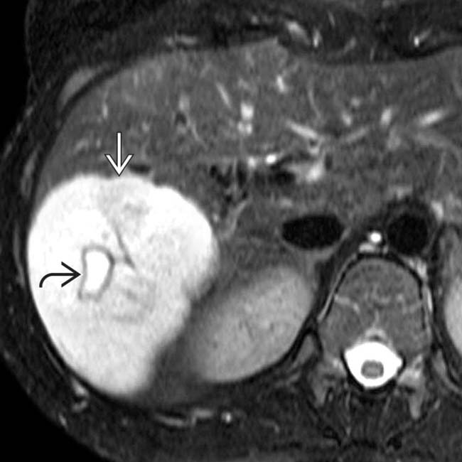
(Left) Axial T2WI MR demonstrates a mass with marked hyperintensity, similar to that of CSF. A central scar within the mass is even more hyperintense, a typical feature of a large or giant hemangioma.
(Right) Axial arterial phase T1WI MR in the same patient shows nodular, discontinuous, peripheral enhancement of the hemangioma, isointense to hepatic vessels, that persisted and progressed on subsequent phases (not shown).
TERMINOLOGY
Synonyms
• Cavernous hemangioma of liver
• Capillary hemangioma (small lesion)
Definitions
• Benign tumor composed of multiple vascular channels lined by single layer of endothelial cells supported by thin fibrous stroma
IMAGING
General Features
• Best diagnostic clue
Peripheral nodular enhancement on arterial phase scan with slow, progressive, centripetal enhancement isodense to vessels
• Location
Common in subcapsular area in posterior right lobe of liver
• Size
Varies from few mm to > 20 cm
Giant hemangiomas: > 10 cm (arbitrary)
• Morphology
Usually solitary and slow growing
May be multiple in up to 50% of cases
Calcification is rare (< 10%)
– Usually within scar of giant hemangioma
CT Findings
• NECT
Small (1-2 cm) and typical (2-10 cm) hemangioma
– Well-circumscribed, spherical to ovoid mass isodense to blood
Giant hemangioma (> 10 cm)
– Heterogeneous hypodense mass
– Central low-density scar ± calcification
• CECT
Small hemangiomas (capillary): < 2 cm
– Arterial and venous phases: Usually show homogeneous enhancement (flash-filling)
Typical hemangiomas: 2-10 cm in diameter
– Arterial phase: Early peripheral, nodular or globular, discontinuous enhancement
– Venous phase: Progressive centripetal enhancement to uniform filling, still isodense to blood vessels
– Delayed phase: Persistent complete filling
Giant hemangioma: > 10 cm in diameter
– Arterial phase: Typical peripheral nodular, cloud-like, or globular enhancement
– Venous and delayed phases: Incomplete centripetal filling of lesion (scar does not enhance)
Atypical hemangioma
– May appear to enhance from inside in centrifugal pattern
– Coronal imaging may reveal more typical centripetal enhancement pattern
Hyalinized (sclerosed) hemangioma
– Shows minimal or no enhancement
– Cannot be diagnosed with confidence by imaging
– Probably the same as “solitary necrotic nodule” described by pathologists
Hemangioma in cirrhotic liver
– Flash-filling of small lesion may mimic hepatocellular carcinoma (HCC)
Does not washout, unlike HCC
– ↓ size and ↑ fibrosis over time
May lose characteristic enhancement pattern
Capsular retraction over shrunken lesion
MR Findings
• T1WI
Small and typical hemangiomas
– Well marginated
– Isointense to blood or hypointense
Giant hemangioma
– Hypointense mass
– Central cleft-like area of marked decreased intensity (scar or fibrous tissue)
• T2WI
Small and typical hemangiomas
– Hyperintense, similar to CSF
Giant hemangioma
– Hyperintense mass
– Marked hyperintense center (scar or fibrosis)
– Hypointense internal septa
• T1WI C+
Same enhancement pattern as on CT
Small hemangiomas (< 2 cm)
– Homogeneous enhancement in arterial and portal phases


 as nonencapsulated collections of blood within enlarged sinusoidal spaces. The liver is otherwise normal.
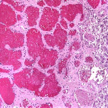
 with marked hyperintensity, similar to that of CSF. A central scar
  within the mass is even more hyperintense, a typical feature of a large or giant hemangioma.
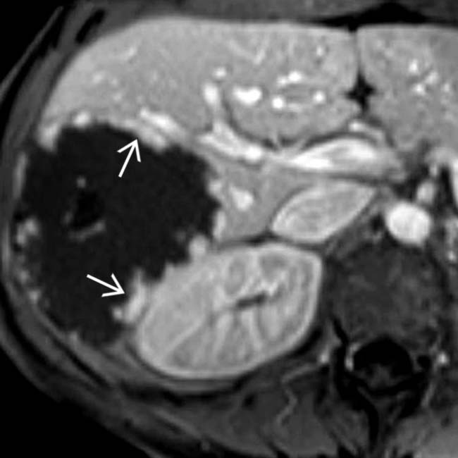
 of the hemangioma, isointense to hepatic vessels, that persisted and progressed on subsequent phases (not shown).


